{"title":"Assessment of intrahepatic cholangiocarcinoma with LI-RADS in the high-risk population: MRI diagnosis and postoperative survival.","authors":"Ruofan Sheng, Beixuan Zheng, Yunfei Zhang, Chun Yang, Dong Wu, Jianjun Zhou, Mengsu Zeng","doi":"10.1186/s40644-025-00860-6","DOIUrl":null,"url":null,"abstract":"<p><strong>Background: </strong>The precise impact of LI-RADS-defined risk factors on the diagnosis and prognosis of intrahepatic cholangiocarcinoma (iCCA) remains unclear.</p><p><strong>Objective: </strong>To assess the value of LI-RADS categories and features for iCCA diagnosis, focusing on the diagnostic and prognostic implications of LI-RADS-defined risk factors.</p><p><strong>Methods: </strong>Totally 214 high risk patients, including 107 surgically-confirmed solitary iCCAs and 107 hepatocellular carcinomas (HCC) from two centers were retrospectively enrolled. Clinical and MRI features based on LI-RADS v2018 were compared, and the performance of targetoid features for discriminating iCCA was evaluated. Recurrence-free survival (RFS) was compared across different pathologic diagnoses and LI-RADS categories. Multivariate Cox analysis was performed to identify the independent risk factors for RFS.</p><p><strong>Results: </strong>In the LI-RADS defined high-risk patients, iCCAs differed from HCCs in MRI manifestation. The LR-M category enabled the accurate classification of most iCCAs (89/107, 83.2%), achieving high sensitivity (83.2%), specificity (85.1%), and accuracy (84.1%). The optimal diagnostic performance for iCCA was achieved when at least one targetoid appearance was required for LR-M categorization (AUC = 0.828). Although 26.2% iCCAs presented at least one major feature and 15.0% iCCAs were miscategorized as probably or definitely HCC, only one iCCA case was categorized as LR-5. RFS varied according to both pathologic diagnosis (P = 0.030) and LI-RADS category (P = 0.028), with LI-RADS category demonstrating an independent association with RFS (HR = 1.736, P = 0.033).</p><p><strong>Conclusions: </strong>In high-risk patients, iCCAs frequently exhibit HCC major features, leading to miscategorization as probable HCC. However, the LR-5 category remains highly specific for ruling out iCCA. Furthermore, in high-risk patients with solitary resected iCCA or HCC, LI-RADS category enables the prediction of postsurgical prognosis independently from pathological diagnosis.</p>","PeriodicalId":9548,"journal":{"name":"Cancer Imaging","volume":"25 1","pages":"40"},"PeriodicalIF":3.5000,"publicationDate":"2025-03-26","publicationTypes":"Journal Article","fieldsOfStudy":null,"isOpenAccess":false,"openAccessPdf":"https://www.ncbi.nlm.nih.gov/pmc/articles/PMC11938583/pdf/","citationCount":"0","resultStr":null,"platform":"Semanticscholar","paperid":null,"PeriodicalName":"Cancer Imaging","FirstCategoryId":"3","ListUrlMain":"https://doi.org/10.1186/s40644-025-00860-6","RegionNum":2,"RegionCategory":"医学","ArticlePicture":[],"TitleCN":null,"AbstractTextCN":null,"PMCID":null,"EPubDate":"","PubModel":"","JCR":"Q2","JCRName":"ONCOLOGY","Score":null,"Total":0}
引用次数: 0
Abstract
Background: The precise impact of LI-RADS-defined risk factors on the diagnosis and prognosis of intrahepatic cholangiocarcinoma (iCCA) remains unclear.
Objective: To assess the value of LI-RADS categories and features for iCCA diagnosis, focusing on the diagnostic and prognostic implications of LI-RADS-defined risk factors.
Methods: Totally 214 high risk patients, including 107 surgically-confirmed solitary iCCAs and 107 hepatocellular carcinomas (HCC) from two centers were retrospectively enrolled. Clinical and MRI features based on LI-RADS v2018 were compared, and the performance of targetoid features for discriminating iCCA was evaluated. Recurrence-free survival (RFS) was compared across different pathologic diagnoses and LI-RADS categories. Multivariate Cox analysis was performed to identify the independent risk factors for RFS.
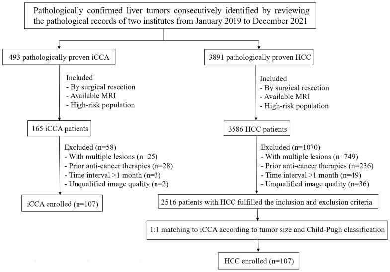
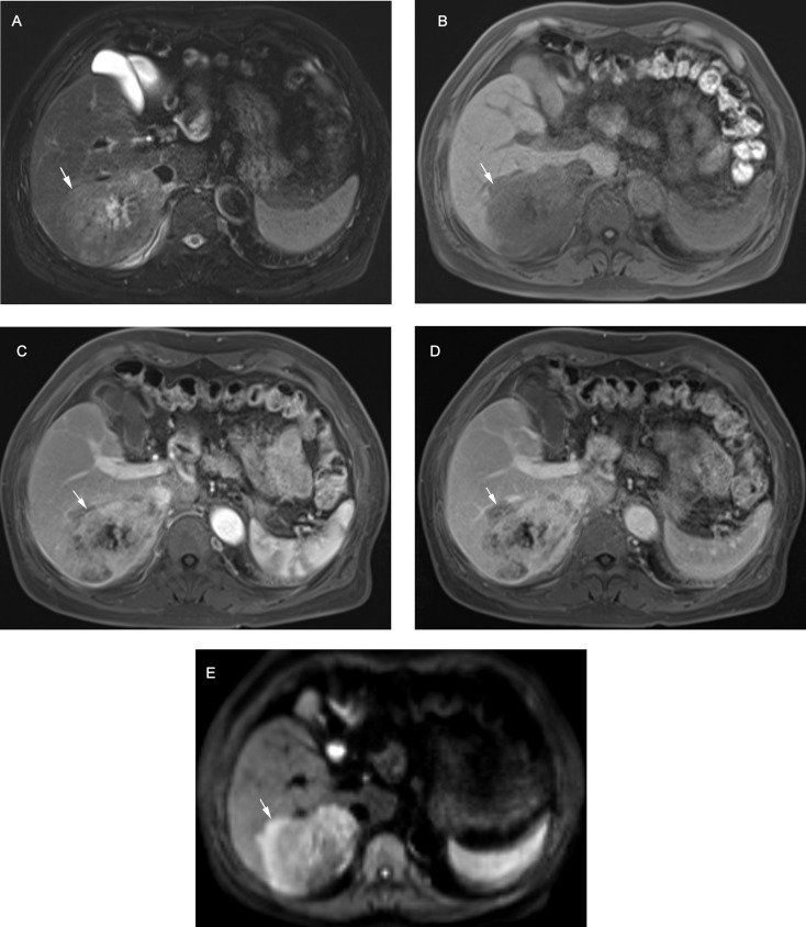
Results: In the LI-RADS defined high-risk patients, iCCAs differed from HCCs in MRI manifestation. The LR-M category enabled the accurate classification of most iCCAs (89/107, 83.2%), achieving high sensitivity (83.2%), specificity (85.1%), and accuracy (84.1%). The optimal diagnostic performance for iCCA was achieved when at least one targetoid appearance was required for LR-M categorization (AUC = 0.828). Although 26.2% iCCAs presented at least one major feature and 15.0% iCCAs were miscategorized as probably or definitely HCC, only one iCCA case was categorized as LR-5. RFS varied according to both pathologic diagnosis (P = 0.030) and LI-RADS category (P = 0.028), with LI-RADS category demonstrating an independent association with RFS (HR = 1.736, P = 0.033).
Conclusions: In high-risk patients, iCCAs frequently exhibit HCC major features, leading to miscategorization as probable HCC. However, the LR-5 category remains highly specific for ruling out iCCA. Furthermore, in high-risk patients with solitary resected iCCA or HCC, LI-RADS category enables the prediction of postsurgical prognosis independently from pathological diagnosis.
Cancer ImagingONCOLOGY-RADIOLOGY, NUCLEAR MEDICINE & MEDICAL IMAGING
CiteScore
7.00
自引率
0.00%
发文量
66
审稿时长
>12 weeks
期刊介绍:
Cancer Imaging is an open access, peer-reviewed journal publishing original articles, reviews and editorials written by expert international radiologists working in oncology.
The journal encompasses CT, MR, PET, ultrasound, radionuclide and multimodal imaging in all kinds of malignant tumours, plus new developments, techniques and innovations. Topics of interest include:
Breast Imaging
Chest
Complications of treatment
Ear, Nose & Throat
Gastrointestinal
Hepatobiliary & Pancreatic
Imaging biomarkers
Interventional
Lymphoma
Measurement of tumour response
Molecular functional imaging
Musculoskeletal
Neuro oncology
Nuclear Medicine
Paediatric.

 求助内容:
求助内容: 应助结果提醒方式:


