Lucas Moreira Mendonça, Marianna Soares Nogueira Borges, Ayodele Alves Amorim, Bruna Neves de Freitas, Camila Tirapelli
{"title":"Kennedy class III and IV dental arches: Trueness analysis of digitization methods and 3D-printing step.","authors":"Lucas Moreira Mendonça, Marianna Soares Nogueira Borges, Ayodele Alves Amorim, Bruna Neves de Freitas, Camila Tirapelli","doi":"10.1590/0103-6440202405980","DOIUrl":null,"url":null,"abstract":"<p><p>This study aims to evaluate the trueness of Kennedy Class III and IV dental arches digitized by different methods and three-dimensionally (3D) printed using stereolithography technology in an in vitro setup. Reference casts (maxillary Kennedy class III and IV) were produced by computer assisted design and manufacture, and linearly measured at occlusocervical, interarch, and edentulous space dimensions. Intraoral scanner (IOS), extraoral scanner (EOS) and cone beam computed tomography (CBCT) digitized the reference casts. Each digital file was 3D-printed using stereolithography technology, totalizing sixty experimental casts (n=10 per group). The same measurements taken from the reference casts were performed on experimental casts. Two-way ANOVA and Bonferroni post-test were used for trueness (distortion between the experimental and reference casts). Distortion was significantly greater for class IV when compared with class III and increased after the 3D-printing step. Among digitizing methods, IOS and EOS had a similar performance and casts from CBCT showed higher distortion, reaching -1.0 and -1.4 mm in the edentulous spaces of digital and 3D-printed cast, respectively. It was possible to conclude that the trueness of Kennedy class III and IV arches were different according to digitizing processes with higher distortion at the edentulous spaces when the cast was digitized by CBCT and converted to a 3D model, compared to IOS and EOS; and in the Kennedy class IV dental arch condition.</p>","PeriodicalId":101363,"journal":{"name":"Brazilian dental journal","volume":"35 ","pages":"e245980"},"PeriodicalIF":0.0000,"publicationDate":"2024-12-06","publicationTypes":"Journal Article","fieldsOfStudy":null,"isOpenAccess":false,"openAccessPdf":"https://www.ncbi.nlm.nih.gov/pmc/articles/PMC11653756/pdf/","citationCount":"0","resultStr":null,"platform":"Semanticscholar","paperid":null,"PeriodicalName":"Brazilian dental journal","FirstCategoryId":"1085","ListUrlMain":"https://doi.org/10.1590/0103-6440202405980","RegionNum":0,"RegionCategory":null,"ArticlePicture":[],"TitleCN":null,"AbstractTextCN":null,"PMCID":null,"EPubDate":"2024/1/1 0:00:00","PubModel":"eCollection","JCR":"","JCRName":"","Score":null,"Total":0}
引用次数: 0
Abstract
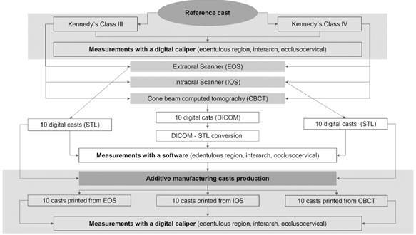
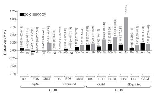
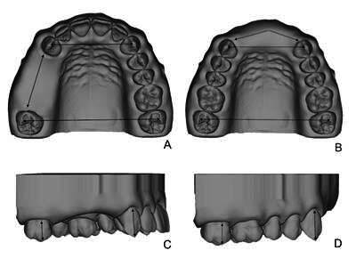
This study aims to evaluate the trueness of Kennedy Class III and IV dental arches digitized by different methods and three-dimensionally (3D) printed using stereolithography technology in an in vitro setup. Reference casts (maxillary Kennedy class III and IV) were produced by computer assisted design and manufacture, and linearly measured at occlusocervical, interarch, and edentulous space dimensions. Intraoral scanner (IOS), extraoral scanner (EOS) and cone beam computed tomography (CBCT) digitized the reference casts. Each digital file was 3D-printed using stereolithography technology, totalizing sixty experimental casts (n=10 per group). The same measurements taken from the reference casts were performed on experimental casts. Two-way ANOVA and Bonferroni post-test were used for trueness (distortion between the experimental and reference casts). Distortion was significantly greater for class IV when compared with class III and increased after the 3D-printing step. Among digitizing methods, IOS and EOS had a similar performance and casts from CBCT showed higher distortion, reaching -1.0 and -1.4 mm in the edentulous spaces of digital and 3D-printed cast, respectively. It was possible to conclude that the trueness of Kennedy class III and IV arches were different according to digitizing processes with higher distortion at the edentulous spaces when the cast was digitized by CBCT and converted to a 3D model, compared to IOS and EOS; and in the Kennedy class IV dental arch condition.

 求助内容:
求助内容: 应助结果提醒方式:


