Mario Henrique Arruda Verzola, Fausto Frizzera, Paulo Sergio Cerri, Ubirajara Pereira Rodrigues-Filho, Silvana Regina Perez Orrico, Rafael Scaf de Molon
{"title":"Effect of Long-Term Treatment with Alendronate on Bone Repair and Mineralization Around Implants in Rats.","authors":"Mario Henrique Arruda Verzola, Fausto Frizzera, Paulo Sergio Cerri, Ubirajara Pereira Rodrigues-Filho, Silvana Regina Perez Orrico, Rafael Scaf de Molon","doi":"10.1590/0103-644020235751","DOIUrl":null,"url":null,"abstract":"<p><p>The aim of this study was to evaluate the effects of long-term alendronate administration on bone repair and mineralization around osseointegrated implants in rats. A total of 160 female Wistar rats were randomly assigned to two groups: the control group (CTL) and the alendronate group (ALD). The ALD group received a subcutaneous injection of sodium alendronate (1 mg/kg/week), while the CTL group received weekly injections of saline solution. After 120 days of treatment, a bilateral implant was placed in the tibia of each rat. Ten rats from each group were euthanized at 5, 10, 15, 20, 25, 30, 45, or 60 days post-surgery. Picro-sirius red staining was utilized to assess the distribution and arrangement of collagen fibers near the implant threads. Bone mineralization mapping of the native bone adjacent to the implant was performed using images obtained through scanning electron microscopy (SEM) across all follow-up periods. SEM-based mineralization mapping revealed an increase in both the degree and homogeneity of bone mineralization in the ALD group compared to the CTL group. Alendronate administration affected collagen arrangement and distribution, leading to a connective tissue with reduced organization and thinner collagen fiber bundles. In conclusion, the findings demonstrated that alendronate administration resulted in a higher degree and homogeneity of bone mineralization, accompanied by reduced collagen content and organization, suggesting an impairment in bone remodeling around dental implants.</p>","PeriodicalId":101363,"journal":{"name":"Brazilian dental journal","volume":"35 ","pages":"e235751"},"PeriodicalIF":0.0000,"publicationDate":"2024-12-06","publicationTypes":"Journal Article","fieldsOfStudy":null,"isOpenAccess":false,"openAccessPdf":"https://www.ncbi.nlm.nih.gov/pmc/articles/PMC11653788/pdf/","citationCount":"0","resultStr":null,"platform":"Semanticscholar","paperid":null,"PeriodicalName":"Brazilian dental journal","FirstCategoryId":"1085","ListUrlMain":"https://doi.org/10.1590/0103-644020235751","RegionNum":0,"RegionCategory":null,"ArticlePicture":[],"TitleCN":null,"AbstractTextCN":null,"PMCID":null,"EPubDate":"2024/1/1 0:00:00","PubModel":"eCollection","JCR":"","JCRName":"","Score":null,"Total":0}
引用次数: 0
Abstract
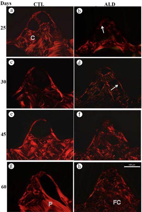
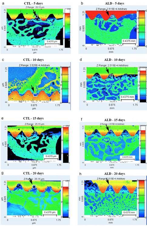
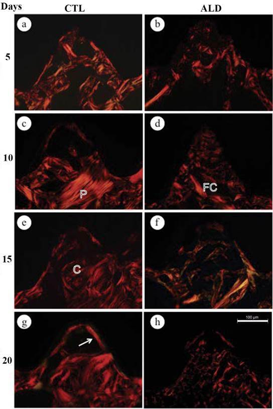
The aim of this study was to evaluate the effects of long-term alendronate administration on bone repair and mineralization around osseointegrated implants in rats. A total of 160 female Wistar rats were randomly assigned to two groups: the control group (CTL) and the alendronate group (ALD). The ALD group received a subcutaneous injection of sodium alendronate (1 mg/kg/week), while the CTL group received weekly injections of saline solution. After 120 days of treatment, a bilateral implant was placed in the tibia of each rat. Ten rats from each group were euthanized at 5, 10, 15, 20, 25, 30, 45, or 60 days post-surgery. Picro-sirius red staining was utilized to assess the distribution and arrangement of collagen fibers near the implant threads. Bone mineralization mapping of the native bone adjacent to the implant was performed using images obtained through scanning electron microscopy (SEM) across all follow-up periods. SEM-based mineralization mapping revealed an increase in both the degree and homogeneity of bone mineralization in the ALD group compared to the CTL group. Alendronate administration affected collagen arrangement and distribution, leading to a connective tissue with reduced organization and thinner collagen fiber bundles. In conclusion, the findings demonstrated that alendronate administration resulted in a higher degree and homogeneity of bone mineralization, accompanied by reduced collagen content and organization, suggesting an impairment in bone remodeling around dental implants.

 求助内容:
求助内容: 应助结果提醒方式:


