Weiqi Wu, Yuan Si, Juan Yang, Liuyan Wen, Jingrong Li
{"title":"Ankyrin repeat domain 1 is dysregulated in keloids and suppresses keloid fibroblast growth, migration, and extracellular matrix deposition.","authors":"Weiqi Wu, Yuan Si, Juan Yang, Liuyan Wen, Jingrong Li","doi":"10.25259/Cytojournal_111_2024","DOIUrl":null,"url":null,"abstract":"<p><strong>Objective: </strong>The etiology and specific pathological mechanisms of keloids remain elusive. Array expression profiling has revealed dysregulation of the transcription cofactor ankyrin repeat domain 1 (ANKRD1) in keloid fibroblasts. The present study focused on examining the expression pattern of ANKRD1 in keloids and assessing its function in human keloid fibroblasts (HKFs).</p><p><strong>Material and methods: </strong>Differential mRNA expression profiles in keloid fibroblasts were investigated by analyzing data from gene expression omnibus (GEO) datasets. Immunohistochemistry assays were performed to verify the expression patterns of ANKRD1 and claudin 11 (CLDN11) in keloid tissue samples. Functional studies were conducted by transfecting HKFs with either a small interfering RNA (siRNA) targeting ANKRD1 (siANKRD1) or ANKRD1-overexpressing plasmids. The functional impact of ANKRD1 was assessed using cell proliferation, flow cytometry, and Transwell migration assays. mRNA expression was evaluated using reverse transcription polymerase chain reaction, and protein expression was determined using Western blotting.</p><p><strong>Results: </strong>Analysis of the GEO series (GSE) GSE44270 revealed eight differentially expressed mRNAs, with ANKRD1 and CLDN11 being the top two downregulated mRNAs. ANKRD1 expression was observed to be lower in keloid tissues than in normal skin tissues, whereas CLDN11 expression showed no significant difference between the two groups. ANKRD1 overexpression suppressed HKF proliferation, migration, and the expression levels of collagen I, fibronectin, matrix metallopeptidase 9, whereas the opposite effects were observed on ANKRD1 knockdown. ANKRD1 did not affect apoptotic cell levels.</p><p><strong>Conclusion: </strong>ANKRD1 is downregulated in keloids and inhibits the growth, migration, and extracellular matrix deposition of keloid fibroblasts. Thus, ANKRD1 may function as a suppressor in keloid formation.</p>","PeriodicalId":49082,"journal":{"name":"Cytojournal","volume":"22 ","pages":"17"},"PeriodicalIF":3.1000,"publicationDate":"2025-02-13","publicationTypes":"Journal Article","fieldsOfStudy":null,"isOpenAccess":false,"openAccessPdf":"https://www.ncbi.nlm.nih.gov/pmc/articles/PMC11932964/pdf/","citationCount":"0","resultStr":null,"platform":"Semanticscholar","paperid":null,"PeriodicalName":"Cytojournal","FirstCategoryId":"3","ListUrlMain":"https://doi.org/10.25259/Cytojournal_111_2024","RegionNum":4,"RegionCategory":"医学","ArticlePicture":[],"TitleCN":null,"AbstractTextCN":null,"PMCID":null,"EPubDate":"2025/1/1 0:00:00","PubModel":"eCollection","JCR":"Q2","JCRName":"PATHOLOGY","Score":null,"Total":0}
引用次数: 0
Abstract
Objective: The etiology and specific pathological mechanisms of keloids remain elusive. Array expression profiling has revealed dysregulation of the transcription cofactor ankyrin repeat domain 1 (ANKRD1) in keloid fibroblasts. The present study focused on examining the expression pattern of ANKRD1 in keloids and assessing its function in human keloid fibroblasts (HKFs).
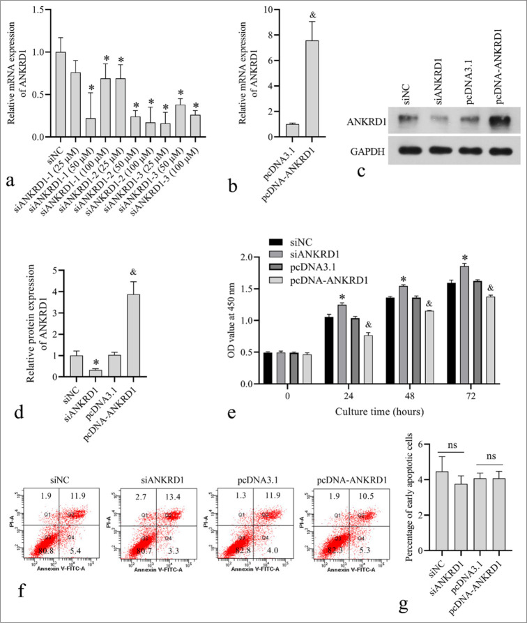
Material and methods: Differential mRNA expression profiles in keloid fibroblasts were investigated by analyzing data from gene expression omnibus (GEO) datasets. Immunohistochemistry assays were performed to verify the expression patterns of ANKRD1 and claudin 11 (CLDN11) in keloid tissue samples. Functional studies were conducted by transfecting HKFs with either a small interfering RNA (siRNA) targeting ANKRD1 (siANKRD1) or ANKRD1-overexpressing plasmids. The functional impact of ANKRD1 was assessed using cell proliferation, flow cytometry, and Transwell migration assays. mRNA expression was evaluated using reverse transcription polymerase chain reaction, and protein expression was determined using Western blotting.
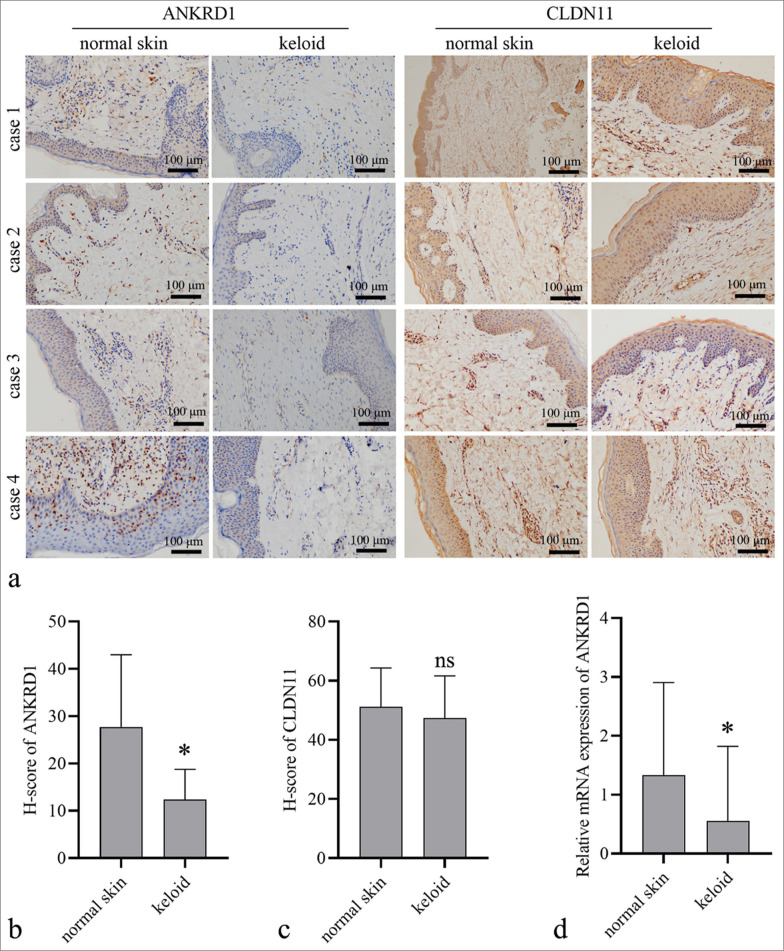
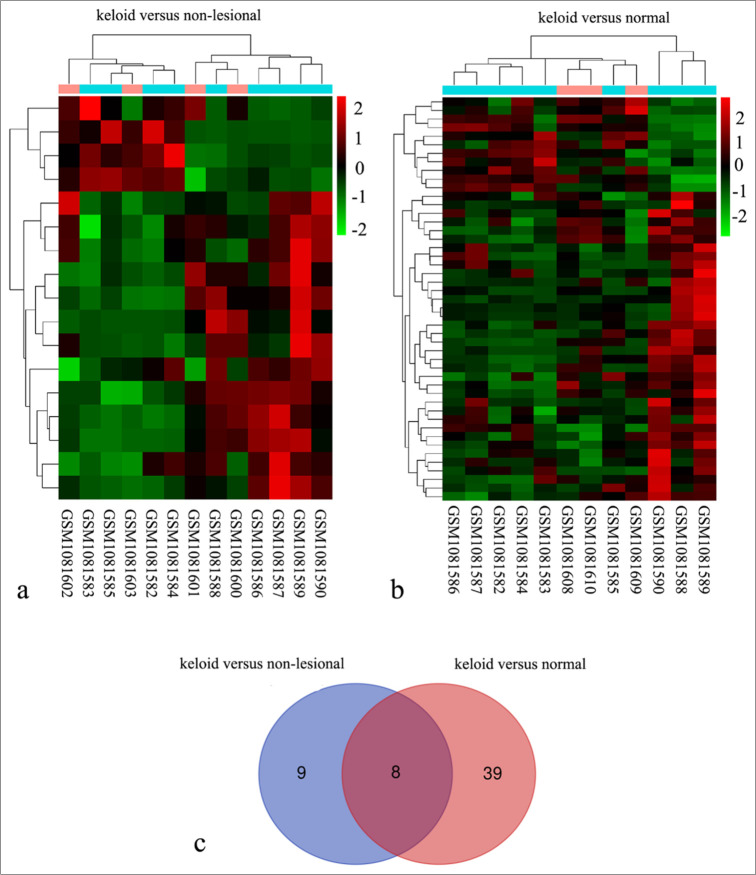
Results: Analysis of the GEO series (GSE) GSE44270 revealed eight differentially expressed mRNAs, with ANKRD1 and CLDN11 being the top two downregulated mRNAs. ANKRD1 expression was observed to be lower in keloid tissues than in normal skin tissues, whereas CLDN11 expression showed no significant difference between the two groups. ANKRD1 overexpression suppressed HKF proliferation, migration, and the expression levels of collagen I, fibronectin, matrix metallopeptidase 9, whereas the opposite effects were observed on ANKRD1 knockdown. ANKRD1 did not affect apoptotic cell levels.
Conclusion: ANKRD1 is downregulated in keloids and inhibits the growth, migration, and extracellular matrix deposition of keloid fibroblasts. Thus, ANKRD1 may function as a suppressor in keloid formation.
期刊介绍:
The CytoJournal is an open-access peer-reviewed journal committed to publishing high-quality articles in the field of Diagnostic Cytopathology including Molecular aspects. The journal is owned by the Cytopathology Foundation and published by the Scientific Scholar.

 求助内容:
求助内容: 应助结果提醒方式:


