Uros Klickovic, Luca Zampedri, Nick Zafeiropoulos, Oliver J Ziff, Christopher Dj Sinclair, Stephen Wastling, Magdalena Dudziec, Jodie Allen, Karin Trimmel, Robin S Howard, Andrea Malaspina, Nikhil Sharma, Katie Cl Sidle, Sachit Shah, Christian Nasel, Tarek A Yousry, Linda Greensmith, Jasper M Morrow, John S Thornton, Pietro Fratta
{"title":"Muscle MRI quantifies disease progression in amyotrophic lateral sclerosis.","authors":"Uros Klickovic, Luca Zampedri, Nick Zafeiropoulos, Oliver J Ziff, Christopher Dj Sinclair, Stephen Wastling, Magdalena Dudziec, Jodie Allen, Karin Trimmel, Robin S Howard, Andrea Malaspina, Nikhil Sharma, Katie Cl Sidle, Sachit Shah, Christian Nasel, Tarek A Yousry, Linda Greensmith, Jasper M Morrow, John S Thornton, Pietro Fratta","doi":"10.1136/jnnp-2024-335571","DOIUrl":null,"url":null,"abstract":"<p><strong>Background and objectives: </strong>Quantitative and operator-independent biomarkers of disease progression are urgently needed in amyotrophic lateral sclerosis (ALS) research. We assess the potential of skeletal muscle MRI as a sensitive and reliable outcome measure for future ALS clinical trials.</p><p><strong>Methods: </strong>In this longitudinal cohort study, muscle MRI of head-neck, upper and lower limb regions, alongside clinical and functional assessments, were acquired at three time points over the individual maximum observation period (iMOP) of 1 year in 20 patients with ALS and 16 healthy controls. Quantitative MRI parameters cross-sectional area (CSA), volume (VOL), fat fraction, functional rest muscle area and water T2 (T<sub>2m</sub>) were correlated with changes in clinical disease severity (functional rating scales and myometry).</p><p><strong>Results: </strong>Among 20 patients with ALS, 17 completed follow-up. Progressive muscle atrophy (CSA, VOL) was observed at hand (rs=0.66), head-neck (partial η²=0.47) and lower-limb level (thighs: η²=0.56, calves: η²=0.54) over iMOP. MRI changes correlated with leg muscle strength (knee extension: r=0.77; plantar flexion: r=0.78), hand grip strength (r=0.71) and functional rating scales (r=0.68).</p><p><strong>Interpretation: </strong>Our findings demonstrate the effectiveness of muscle MRI as a sensitive neuroimaging biomarker of disease progression in ALS, highlighting its potential application in clinical trials.</p>","PeriodicalId":16418,"journal":{"name":"Journal of Neurology, Neurosurgery, and Psychiatry","volume":" ","pages":"908-911"},"PeriodicalIF":7.5000,"publicationDate":"2025-08-14","publicationTypes":"Journal Article","fieldsOfStudy":null,"isOpenAccess":false,"openAccessPdf":"https://www.ncbi.nlm.nih.gov/pmc/articles/PMC12418527/pdf/","citationCount":"0","resultStr":null,"platform":"Semanticscholar","paperid":null,"PeriodicalName":"Journal of Neurology, Neurosurgery, and Psychiatry","FirstCategoryId":"3","ListUrlMain":"https://doi.org/10.1136/jnnp-2024-335571","RegionNum":1,"RegionCategory":"医学","ArticlePicture":[],"TitleCN":null,"AbstractTextCN":null,"PMCID":null,"EPubDate":"","PubModel":"","JCR":"Q1","JCRName":"CLINICAL NEUROLOGY","Score":null,"Total":0}
引用次数: 0
Abstract
Background and objectives: Quantitative and operator-independent biomarkers of disease progression are urgently needed in amyotrophic lateral sclerosis (ALS) research. We assess the potential of skeletal muscle MRI as a sensitive and reliable outcome measure for future ALS clinical trials.
Methods: In this longitudinal cohort study, muscle MRI of head-neck, upper and lower limb regions, alongside clinical and functional assessments, were acquired at three time points over the individual maximum observation period (iMOP) of 1 year in 20 patients with ALS and 16 healthy controls. Quantitative MRI parameters cross-sectional area (CSA), volume (VOL), fat fraction, functional rest muscle area and water T2 (T2m) were correlated with changes in clinical disease severity (functional rating scales and myometry).
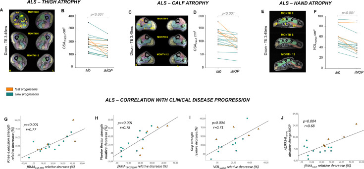
Results: Among 20 patients with ALS, 17 completed follow-up. Progressive muscle atrophy (CSA, VOL) was observed at hand (rs=0.66), head-neck (partial η²=0.47) and lower-limb level (thighs: η²=0.56, calves: η²=0.54) over iMOP. MRI changes correlated with leg muscle strength (knee extension: r=0.77; plantar flexion: r=0.78), hand grip strength (r=0.71) and functional rating scales (r=0.68).
Interpretation: Our findings demonstrate the effectiveness of muscle MRI as a sensitive neuroimaging biomarker of disease progression in ALS, highlighting its potential application in clinical trials.
期刊介绍:
The Journal of Neurology, Neurosurgery & Psychiatry (JNNP) aspires to publish groundbreaking and cutting-edge research worldwide. Covering the entire spectrum of neurological sciences, the journal focuses on common disorders like stroke, multiple sclerosis, Parkinson’s disease, epilepsy, peripheral neuropathy, subarachnoid haemorrhage, and neuropsychiatry, while also addressing complex challenges such as ALS. With early online publication, regular podcasts, and an extensive archive collection boasting the longest half-life in clinical neuroscience journals, JNNP aims to be a trailblazer in the field.
 求助内容:
求助内容: 应助结果提醒方式:


