{"title":"Longitudinal changes in the position and thickness of the peak peripapillary retinal nerve fiber layer in school children.","authors":"Takehiro Yamashita, Hiroto Terasaki, Ryo Asaoka, Naoya Yoshihara, Naoko Kakiuchi, Taiji Sakamoto","doi":"10.1007/s00417-025-06810-z","DOIUrl":null,"url":null,"abstract":"<p><strong>Purpose: </strong>This study investigated the relationship between changes in the position and thickness of the peak circumpapillary retinal nerve fiber layer (cpRNFL) and axial elongation in schoolchildren.</p><p><strong>Methods: </strong>This prospective cohort study involved the right eyes of 75 elementary school students examined over a period of six years (from the age of 8-9 years to 14-15 years). During the first and final years, all participants underwent optical axial length measurements, color fundus photography, and cpRNFL thickness measurements using optical coherence tomography. The supratemporal (ST) and infratemporal (IT) peak angles (ST and IT angle) were defined as those formed by the ST/IT peak position of the cpRNFL curve, the center of the optic disc, and the fovea. The RNFL thickness at the peaks (ST and IT thicknesses) was also determined. The Wilcoxon signed-rank test was used to compare the cpRNFL parameters and axial lengths in the first and final years.</p><p><strong>Results: </strong>The mean axial length was significantly longer in the final year (24.82 mm) than in the first year (23.34 mm). The mean ST and IT angles were significantly lower in the final year (67.6° and 58.2°) than in the first year (74.2° and 64.0°). The mean IT thickness was significantly greater in the final year (195.1 μm) than in the first year (185.0 μm); however, no significant changes in ST thickness were observed.</p><p><strong>Conclusion: </strong>The ST and IT peaks shifted toward the line connecting the fovea and the center of the optic disc between ages 8-9 and 14-15 years, and IT thickness increased. These changes indicate that nerve fibers are concentrated on the temporal side of the optic disc, especially in the IT area.</p><p><strong>Key messages: </strong>WHAT IS KNOWN : The circumpapillary retinal nerve fiber layer (cpRNFL) in normal eyes exhibits a double-hump pattern, with individual variability in the position of the peaks. Additionally, the mechanisms underlying these differences remain unclear.</p><p><strong>What is new: </strong>Eyes with greater axial elongation tended to have narrower supratemporal (ST) and infratemporal (IT) angles and increased IT thickness. Greater axial elongation during childhood growth caused a significant shift of the cpRNFL peaks toward the fovea and increased IT thickness. Based on the plate hypothesis, the shift and compression of nerve fibers during growth may serve as a potential predictor of normal-tension glaucoma onset in the future.</p>","PeriodicalId":12795,"journal":{"name":"Graefe’s Archive for Clinical and Experimental Ophthalmology","volume":" ","pages":"1977-1984"},"PeriodicalIF":2.4000,"publicationDate":"2025-07-01","publicationTypes":"Journal Article","fieldsOfStudy":null,"isOpenAccess":false,"openAccessPdf":"https://www.ncbi.nlm.nih.gov/pmc/articles/PMC12373550/pdf/","citationCount":"0","resultStr":null,"platform":"Semanticscholar","paperid":null,"PeriodicalName":"Graefe’s Archive for Clinical and Experimental Ophthalmology","FirstCategoryId":"3","ListUrlMain":"https://doi.org/10.1007/s00417-025-06810-z","RegionNum":3,"RegionCategory":"医学","ArticlePicture":[],"TitleCN":null,"AbstractTextCN":null,"PMCID":null,"EPubDate":"2025/3/25 0:00:00","PubModel":"Epub","JCR":"Q2","JCRName":"OPHTHALMOLOGY","Score":null,"Total":0}
引用次数: 0
Abstract
Purpose: This study investigated the relationship between changes in the position and thickness of the peak circumpapillary retinal nerve fiber layer (cpRNFL) and axial elongation in schoolchildren.
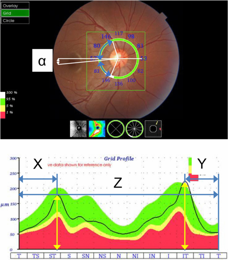
Methods: This prospective cohort study involved the right eyes of 75 elementary school students examined over a period of six years (from the age of 8-9 years to 14-15 years). During the first and final years, all participants underwent optical axial length measurements, color fundus photography, and cpRNFL thickness measurements using optical coherence tomography. The supratemporal (ST) and infratemporal (IT) peak angles (ST and IT angle) were defined as those formed by the ST/IT peak position of the cpRNFL curve, the center of the optic disc, and the fovea. The RNFL thickness at the peaks (ST and IT thicknesses) was also determined. The Wilcoxon signed-rank test was used to compare the cpRNFL parameters and axial lengths in the first and final years.
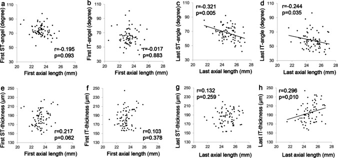
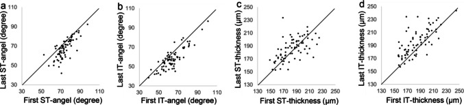
Results: The mean axial length was significantly longer in the final year (24.82 mm) than in the first year (23.34 mm). The mean ST and IT angles were significantly lower in the final year (67.6° and 58.2°) than in the first year (74.2° and 64.0°). The mean IT thickness was significantly greater in the final year (195.1 μm) than in the first year (185.0 μm); however, no significant changes in ST thickness were observed.
Conclusion: The ST and IT peaks shifted toward the line connecting the fovea and the center of the optic disc between ages 8-9 and 14-15 years, and IT thickness increased. These changes indicate that nerve fibers are concentrated on the temporal side of the optic disc, especially in the IT area.
Key messages: WHAT IS KNOWN : The circumpapillary retinal nerve fiber layer (cpRNFL) in normal eyes exhibits a double-hump pattern, with individual variability in the position of the peaks. Additionally, the mechanisms underlying these differences remain unclear.
What is new: Eyes with greater axial elongation tended to have narrower supratemporal (ST) and infratemporal (IT) angles and increased IT thickness. Greater axial elongation during childhood growth caused a significant shift of the cpRNFL peaks toward the fovea and increased IT thickness. Based on the plate hypothesis, the shift and compression of nerve fibers during growth may serve as a potential predictor of normal-tension glaucoma onset in the future.
期刊介绍:
Graefe''s Archive for Clinical and Experimental Ophthalmology is a distinguished international journal that presents original clinical reports and clini-cally relevant experimental studies. Founded in 1854 by Albrecht von Graefe to serve as a source of useful clinical information and a stimulus for discussion, the journal has published articles by leading ophthalmologists and vision research scientists for more than a century. With peer review by an international Editorial Board and prompt English-language publication, Graefe''s Archive provides rapid dissemination of clinical and clinically related experimental information.

 求助内容:
求助内容: 应助结果提醒方式:


