Mecit Kantarcı, Volkan Kızılgöz, Ramazan Terzi, Ahmet Enes Kılıç, Halime Kabalcı, Önder Durmaz, Nil Tokgöz, Mustafa Harman, Ayşegül Sağır Kahraman, Ali Avanaz, Sonay Aydın, Gülsüm Özlem Elpek, Merve Yazol, Bülent Aydınlı
{"title":"Evaluating artificial intelligence for a focal nodular hyperplasia diagnosis using magnetic resonance imaging: preliminary findings.","authors":"Mecit Kantarcı, Volkan Kızılgöz, Ramazan Terzi, Ahmet Enes Kılıç, Halime Kabalcı, Önder Durmaz, Nil Tokgöz, Mustafa Harman, Ayşegül Sağır Kahraman, Ali Avanaz, Sonay Aydın, Gülsüm Özlem Elpek, Merve Yazol, Bülent Aydınlı","doi":"10.4274/dir.2025.243095","DOIUrl":null,"url":null,"abstract":"<p><strong>Purpose: </strong>This study aimed to evaluate the effectiveness of artificial intelligence (AI) in diagnosing focal nodular hyperplasia (FNH) of the liver using magnetic resonance imaging (MRI) and compare its performance with that of radiologists.</p><p><strong>Methods: </strong>In the first phase of the study, the MRIs of 60 patients (30 patients with FNH and 30 patients with no lesions or lesions other than FNH) were processed using a segmentation program and introduced to an AI model. After the learning process, the MRIs of 42 different patients that the AI model had no experience with were introduced to the system. In addition, a radiology resident and a radiology specialist evaluated patients with the same MR sequences. The sensitivity and specificity values were obtained from all three reviews.</p><p><strong>Results: </strong>The sensitivity, specificity, positive predictive value (PPV), and negative predictive value (NPV) of the AI model were found to be 0.769, 0.966, 0.909, and 0.903, respectively. The sensitivity and specificity values were higher than those of the radiology resident and lower than those of the radiology specialist. The results of the specialist versus the AI model revealed a good agreement level, with a kappa (κ) value of 0.777.</p><p><strong>Conclusion: </strong>For the diagnosis of FNH, the sensitivity, specificity, PPV, and NPV of the AI device were higher than those of the radiology resident and lower than those of the radiology specialist. With additional studies focused on different specific lesions of the liver, AI models are expected to be able to diagnose each liver lesion with high accuracy in the future.</p><p><strong>Clinical significance: </strong>AI is studied to provide assisted or automated interpretation of radiological images with an accurate and reproducible imaging diagnosis.</p>","PeriodicalId":11341,"journal":{"name":"Diagnostic and interventional radiology","volume":" ","pages":"405-415"},"PeriodicalIF":1.7000,"publicationDate":"2025-09-08","publicationTypes":"Journal Article","fieldsOfStudy":null,"isOpenAccess":false,"openAccessPdf":"https://www.ncbi.nlm.nih.gov/pmc/articles/PMC12417915/pdf/","citationCount":"0","resultStr":null,"platform":"Semanticscholar","paperid":null,"PeriodicalName":"Diagnostic and interventional radiology","FirstCategoryId":"3","ListUrlMain":"https://doi.org/10.4274/dir.2025.243095","RegionNum":4,"RegionCategory":"医学","ArticlePicture":[],"TitleCN":null,"AbstractTextCN":null,"PMCID":null,"EPubDate":"2025/3/26 0:00:00","PubModel":"Epub","JCR":"Q3","JCRName":"RADIOLOGY, NUCLEAR MEDICINE & MEDICAL IMAGING","Score":null,"Total":0}
引用次数: 0
Abstract
Purpose: This study aimed to evaluate the effectiveness of artificial intelligence (AI) in diagnosing focal nodular hyperplasia (FNH) of the liver using magnetic resonance imaging (MRI) and compare its performance with that of radiologists.
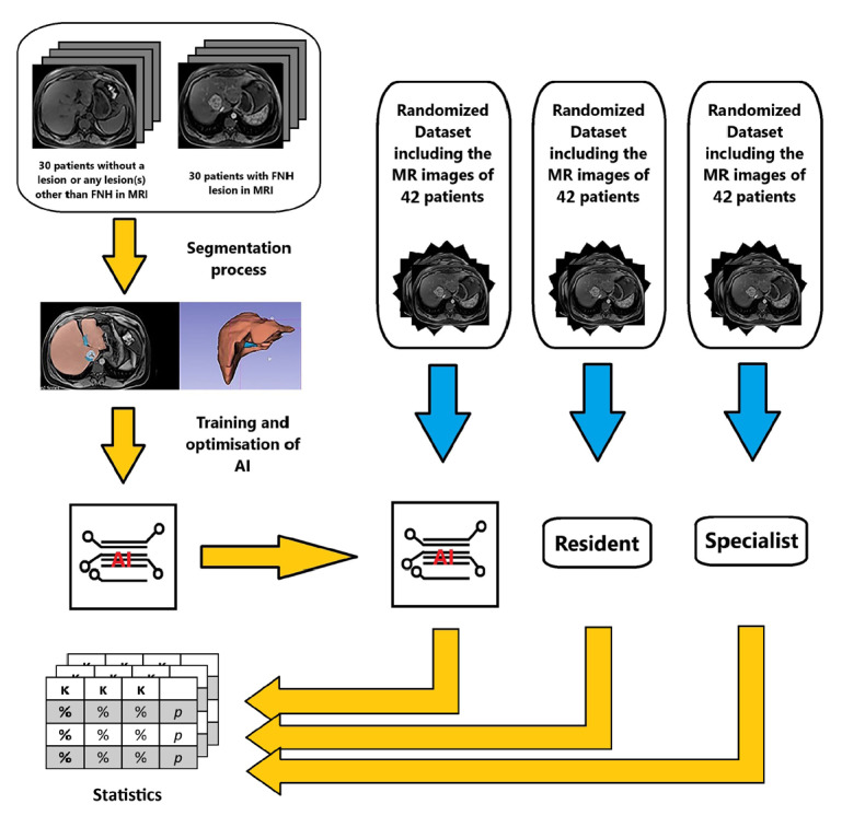
Methods: In the first phase of the study, the MRIs of 60 patients (30 patients with FNH and 30 patients with no lesions or lesions other than FNH) were processed using a segmentation program and introduced to an AI model. After the learning process, the MRIs of 42 different patients that the AI model had no experience with were introduced to the system. In addition, a radiology resident and a radiology specialist evaluated patients with the same MR sequences. The sensitivity and specificity values were obtained from all three reviews.
Results: The sensitivity, specificity, positive predictive value (PPV), and negative predictive value (NPV) of the AI model were found to be 0.769, 0.966, 0.909, and 0.903, respectively. The sensitivity and specificity values were higher than those of the radiology resident and lower than those of the radiology specialist. The results of the specialist versus the AI model revealed a good agreement level, with a kappa (κ) value of 0.777.
Conclusion: For the diagnosis of FNH, the sensitivity, specificity, PPV, and NPV of the AI device were higher than those of the radiology resident and lower than those of the radiology specialist. With additional studies focused on different specific lesions of the liver, AI models are expected to be able to diagnose each liver lesion with high accuracy in the future.
Clinical significance: AI is studied to provide assisted or automated interpretation of radiological images with an accurate and reproducible imaging diagnosis.
期刊介绍:
Diagnostic and Interventional Radiology (Diagn Interv Radiol) is the open access, online-only official publication of Turkish Society of Radiology. It is published bimonthly and the journal’s publication language is English.
The journal is a medium for original articles, reviews, pictorial essays, technical notes related to all fields of diagnostic and interventional radiology.

 求助内容:
求助内容: 应助结果提醒方式:


