Mohammed A Azab, Ahmed Hazim, Nour El-Gohary, Mohsen Nabih Shama, Brahim Kammoun
{"title":"Calvarial Chondroplastic Osteosarcoma With Distant Brain Metastasis Treated With Radiosurgery: A Rare Case Report.","authors":"Mohammed A Azab, Ahmed Hazim, Nour El-Gohary, Mohsen Nabih Shama, Brahim Kammoun","doi":"10.1155/carm/5412921","DOIUrl":null,"url":null,"abstract":"<p><p><b>Background:</b> Cerebral metastases from soft tissue and bone sarcoma are uncommon. Metastatic sarcoma of the brain is a highly aggressive disease with a poor prognosis. There is no consensus regarding the management of cerebral metastases from bone sarcomas. <b>Clinical Presentation:</b> The patient is a 60-year-old, right-handed male, who presented with a right frontal scalp swelling that was hard in consistency. On examination, he had pain and tenderness over the swelling. The neurological examination was normal. <b>Investigations:</b> Initial CTH revealed a right frontal skull lesion with characteristic expansion and sunburst appearance with a degree of cortical destruction. MRI brain with contrast showed features suggestive of skull osteosarcoma. <b>Management:</b> He underwent a subtotal tumor resection. He was diagnosed with high-grade chondroblastoma-like osteosarcoma of the skull. Subsequently, he received three cycles of neoadjuvant chemotherapy in the form of Adriamycin and cisplatin. One year later, he underwent further surgical intervention with an additional skull resection and reconstruction using mesh and scalp reconstruction. <b>Follow-Up:</b> MRI brain with contrast showed a distant metastasis in the right transverse sinus and other distant brain areas and were treated with Gamma Knife radiosurgery (GKRS) 6 months after the primary surgery. <b>Conclusion:</b> Skull calvarium primary osteosarcoma is a rare pathology. Cerebral metastasis from skull bone osteosarcoma is a challenging clinical situation that requires a multidisciplinary therapeutic approach that includes neurosurgery, plastic surgery, chemotherapy, and radiosurgery.</p>","PeriodicalId":9627,"journal":{"name":"Case Reports in Medicine","volume":"2025 ","pages":"5412921"},"PeriodicalIF":0.7000,"publicationDate":"2025-03-18","publicationTypes":"Journal Article","fieldsOfStudy":null,"isOpenAccess":false,"openAccessPdf":"https://www.ncbi.nlm.nih.gov/pmc/articles/PMC11936537/pdf/","citationCount":"0","resultStr":null,"platform":"Semanticscholar","paperid":null,"PeriodicalName":"Case Reports in Medicine","FirstCategoryId":"1085","ListUrlMain":"https://doi.org/10.1155/carm/5412921","RegionNum":0,"RegionCategory":null,"ArticlePicture":[],"TitleCN":null,"AbstractTextCN":null,"PMCID":null,"EPubDate":"2025/1/1 0:00:00","PubModel":"eCollection","JCR":"Q3","JCRName":"MEDICINE, GENERAL & INTERNAL","Score":null,"Total":0}
引用次数: 0
Abstract
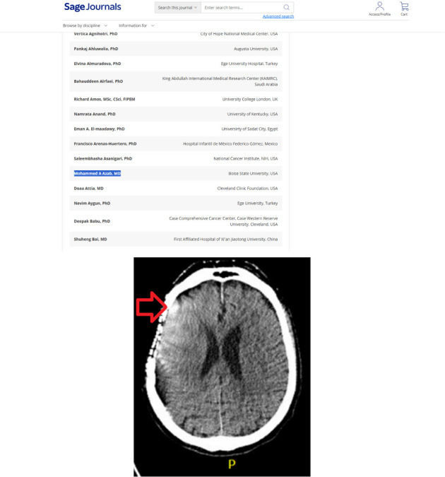
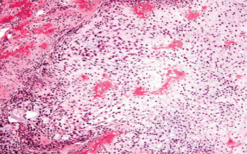
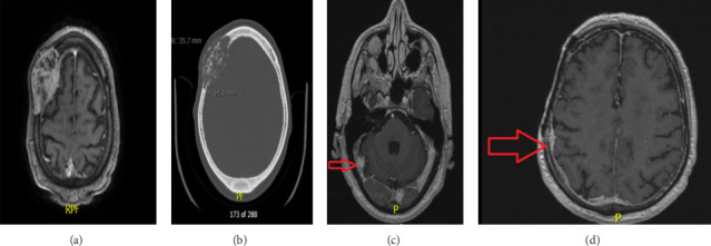
Background: Cerebral metastases from soft tissue and bone sarcoma are uncommon. Metastatic sarcoma of the brain is a highly aggressive disease with a poor prognosis. There is no consensus regarding the management of cerebral metastases from bone sarcomas. Clinical Presentation: The patient is a 60-year-old, right-handed male, who presented with a right frontal scalp swelling that was hard in consistency. On examination, he had pain and tenderness over the swelling. The neurological examination was normal. Investigations: Initial CTH revealed a right frontal skull lesion with characteristic expansion and sunburst appearance with a degree of cortical destruction. MRI brain with contrast showed features suggestive of skull osteosarcoma. Management: He underwent a subtotal tumor resection. He was diagnosed with high-grade chondroblastoma-like osteosarcoma of the skull. Subsequently, he received three cycles of neoadjuvant chemotherapy in the form of Adriamycin and cisplatin. One year later, he underwent further surgical intervention with an additional skull resection and reconstruction using mesh and scalp reconstruction. Follow-Up: MRI brain with contrast showed a distant metastasis in the right transverse sinus and other distant brain areas and were treated with Gamma Knife radiosurgery (GKRS) 6 months after the primary surgery. Conclusion: Skull calvarium primary osteosarcoma is a rare pathology. Cerebral metastasis from skull bone osteosarcoma is a challenging clinical situation that requires a multidisciplinary therapeutic approach that includes neurosurgery, plastic surgery, chemotherapy, and radiosurgery.

 求助内容:
求助内容: 应助结果提醒方式:


