Elizabeth M Teasell, Emilie Potts, Nicole Geremia, Lihong Lu, Xiaoyun Xu, Haojie Mao, Arthur Brown
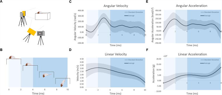
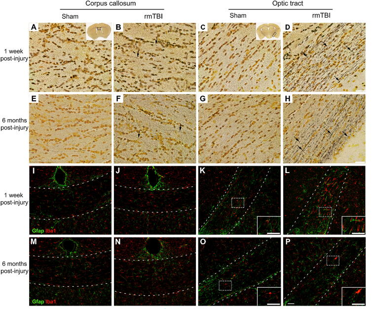
{"title":"A Clinically Relevant Mouse Model of Concussion Incorporating High Rotational Forces.","authors":"Elizabeth M Teasell, Emilie Potts, Nicole Geremia, Lihong Lu, Xiaoyun Xu, Haojie Mao, Arthur Brown","doi":"10.1089/neur.2024.0165","DOIUrl":null,"url":null,"abstract":"<p><p>Clinically relevant models of concussion are critical in understanding the pathophysiology of concussion and its long-term outcomes. To bridge the gap between preclinical and clinical research, animal models of concussion should be produced by mild traumatic brain injuries (mTBIs) that possess the same physical and biomechanical properties found in the mTBIs that cause concussion in humans. Specifically, to have good construct validity the mTBIs used in animal models of concussion should feature closed-head impacts with unrestrained head and body motion, resulting in peak angular velocities that approximate the human experience. We describe a mouse model of concussion using a cortical impactor to deliver closed-head mTBIs. Mice are placed on a break-away platform that allows free head and body movement during and after impact resulting in rapid head rotation. We assessed this model of concussion in over 100 mice carrying humanized versions of the genes encoding the amyloid precursor protein and tau. We found that this method consistently produced injuries with peak angular velocities in mice that, when scaled, approximated the average peak angular velocities reported in concussive football impacts. Face validity of this model of concussion was evaluated by histopathology and revealed that three impacts delivered 24 hours apart led to diffuse axonal injury, astrogliosis, and microglial activation one week after injury, particularly in white matter tracts aligned orthogonally to the axis of rotation. Persistent axonal degeneration was observed up to 6 months postinjury. This mouse model of concussion captures key biomechanical and pathological features of human concussions.</p>","PeriodicalId":74300,"journal":{"name":"Neurotrauma reports","volume":"6 1","pages":"184-190"},"PeriodicalIF":1.8000,"publicationDate":"2025-02-17","publicationTypes":"Journal Article","fieldsOfStudy":null,"isOpenAccess":false,"openAccessPdf":"https://www.ncbi.nlm.nih.gov/pmc/articles/PMC11931110/pdf/","citationCount":"0","resultStr":null,"platform":"Semanticscholar","paperid":null,"PeriodicalName":"Neurotrauma reports","FirstCategoryId":"1085","ListUrlMain":"https://doi.org/10.1089/neur.2024.0165","RegionNum":0,"RegionCategory":null,"ArticlePicture":[],"TitleCN":null,"AbstractTextCN":null,"PMCID":null,"EPubDate":"2025/1/1 0:00:00","PubModel":"eCollection","JCR":"Q3","JCRName":"CLINICAL NEUROLOGY","Score":null,"Total":0}
引用次数: 0
Abstract
Clinically relevant models of concussion are critical in understanding the pathophysiology of concussion and its long-term outcomes. To bridge the gap between preclinical and clinical research, animal models of concussion should be produced by mild traumatic brain injuries (mTBIs) that possess the same physical and biomechanical properties found in the mTBIs that cause concussion in humans. Specifically, to have good construct validity the mTBIs used in animal models of concussion should feature closed-head impacts with unrestrained head and body motion, resulting in peak angular velocities that approximate the human experience. We describe a mouse model of concussion using a cortical impactor to deliver closed-head mTBIs. Mice are placed on a break-away platform that allows free head and body movement during and after impact resulting in rapid head rotation. We assessed this model of concussion in over 100 mice carrying humanized versions of the genes encoding the amyloid precursor protein and tau. We found that this method consistently produced injuries with peak angular velocities in mice that, when scaled, approximated the average peak angular velocities reported in concussive football impacts. Face validity of this model of concussion was evaluated by histopathology and revealed that three impacts delivered 24 hours apart led to diffuse axonal injury, astrogliosis, and microglial activation one week after injury, particularly in white matter tracts aligned orthogonally to the axis of rotation. Persistent axonal degeneration was observed up to 6 months postinjury. This mouse model of concussion captures key biomechanical and pathological features of human concussions.

 求助内容:
求助内容: 应助结果提醒方式:


