Konstantin Klambauer, Thomas Flohr, Lukas Jakob Moser, Victor Mergen, Matthias Eberhard, Andreas Prokein, Hatem Alkadhi, Hubertus Pietsch, Gregor Jost
{"title":"Reducing contrast media and radiation dose in CT angiography at low tube voltage: animal study with photon-counting detector CT.","authors":"Konstantin Klambauer, Thomas Flohr, Lukas Jakob Moser, Victor Mergen, Matthias Eberhard, Andreas Prokein, Hatem Alkadhi, Hubertus Pietsch, Gregor Jost","doi":"10.1186/s41747-025-00577-y","DOIUrl":null,"url":null,"abstract":"<p><strong>Background: </strong>Reducing radiation and contrast media (CM) doses in computed tomography angiography (CTA) is especially relevant for potentially vulnerable populations. Low tube voltage photon-counting detector CT (PCD-CT) offers an improved iodine contrast-to-noise ratio (CNR) as compared to conventional CT scanners. We investigated optimized radiation and CM doses of PCD-CT angiography at low tube voltage in an animal model.</p><p><strong>Methods: </strong>Six minipigs (median weight: 32.5 kg; IQR: 29.8-34.6 kg) underwent thoracoabdominal CTA using a clinical dual-source PCD-CT at 70 kVp with three scan protocols: (A) reference (100% CM and radiation dose), (B) increased radiation (233%) and reduced CM (56%) dose, and (C) reduced radiation (50%) and increased CM (141%) dose. CNR, subjective image quality, and radiation doses were assessed, with statistical analysis including Mann-Whitney U-test and Kruskal-Wallis tests.</p><p><strong>Results: </strong>CTDI<sub>vol</sub> was 1.7 mGy (IQR: 1.5-1.8) for scan A, 4.3 mGy (IQR: 3.8-4.7) for scan B, and 0.9 mGy (IQR: 0.8-1.0) for scan C (p < 0.001). CM volumes were 16 mL (IQR: 15-17) for scan A, 10 mL (IQR: 8-10) for scan B, and 23 mL (IQR: 21-24) for scan C. No significant differences in CNR were found between scans, with medians of 26 (IQR: 24-28) for scan A, 23 (IQR: 22-26) for scan B, and 26 (IQR: 24-30) for scan C (p = 0.276). Subjective image quality was similar across scans (p = 0.342).</p><p><strong>Conclusion: </strong>Low tube voltage PCD-CT angiography allows substantial reductions in radiation and CM dose while maintaining stable and improved CNR, which allows further dose flexibility for individualized CTA protocols.</p><p><strong>Relevance statement: </strong>PCD-CT at low tube voltage provides a high CNR and great flexibility in dose optimization, making it particularly effective for applications where minimizing radiation and CM exposure is a priority.</p><p><strong>Key points: </strong>Low tube voltage imaging with photon counting detector (PCD)-CT enables flexible contrast and radiation dose optimization strategies in thoracoabdominal CT angiography (CTA). The CNR for thoracoabdominal CTA remains stable with appropriate contrast and radiation dose adjustments at low tube voltage PCD-CT. Low tube voltage PCD-CT consistently yields diagnostic image quality in thoracoabdominal angiography even at reduced contrast or radiation doses.</p>","PeriodicalId":36926,"journal":{"name":"European Radiology Experimental","volume":"9 1","pages":"37"},"PeriodicalIF":3.6000,"publicationDate":"2025-03-24","publicationTypes":"Journal Article","fieldsOfStudy":null,"isOpenAccess":false,"openAccessPdf":"https://www.ncbi.nlm.nih.gov/pmc/articles/PMC11933636/pdf/","citationCount":"0","resultStr":null,"platform":"Semanticscholar","paperid":null,"PeriodicalName":"European Radiology Experimental","FirstCategoryId":"1085","ListUrlMain":"https://doi.org/10.1186/s41747-025-00577-y","RegionNum":0,"RegionCategory":null,"ArticlePicture":[],"TitleCN":null,"AbstractTextCN":null,"PMCID":null,"EPubDate":"","PubModel":"","JCR":"Q1","JCRName":"RADIOLOGY, NUCLEAR MEDICINE & MEDICAL IMAGING","Score":null,"Total":0}
引用次数: 0
Abstract
Background: Reducing radiation and contrast media (CM) doses in computed tomography angiography (CTA) is especially relevant for potentially vulnerable populations. Low tube voltage photon-counting detector CT (PCD-CT) offers an improved iodine contrast-to-noise ratio (CNR) as compared to conventional CT scanners. We investigated optimized radiation and CM doses of PCD-CT angiography at low tube voltage in an animal model.
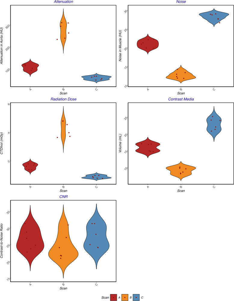
Methods: Six minipigs (median weight: 32.5 kg; IQR: 29.8-34.6 kg) underwent thoracoabdominal CTA using a clinical dual-source PCD-CT at 70 kVp with three scan protocols: (A) reference (100% CM and radiation dose), (B) increased radiation (233%) and reduced CM (56%) dose, and (C) reduced radiation (50%) and increased CM (141%) dose. CNR, subjective image quality, and radiation doses were assessed, with statistical analysis including Mann-Whitney U-test and Kruskal-Wallis tests.
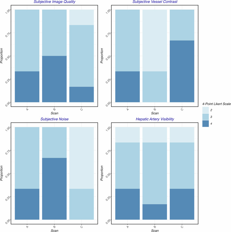
Results: CTDIvol was 1.7 mGy (IQR: 1.5-1.8) for scan A, 4.3 mGy (IQR: 3.8-4.7) for scan B, and 0.9 mGy (IQR: 0.8-1.0) for scan C (p < 0.001). CM volumes were 16 mL (IQR: 15-17) for scan A, 10 mL (IQR: 8-10) for scan B, and 23 mL (IQR: 21-24) for scan C. No significant differences in CNR were found between scans, with medians of 26 (IQR: 24-28) for scan A, 23 (IQR: 22-26) for scan B, and 26 (IQR: 24-30) for scan C (p = 0.276). Subjective image quality was similar across scans (p = 0.342).
Conclusion: Low tube voltage PCD-CT angiography allows substantial reductions in radiation and CM dose while maintaining stable and improved CNR, which allows further dose flexibility for individualized CTA protocols.
Relevance statement: PCD-CT at low tube voltage provides a high CNR and great flexibility in dose optimization, making it particularly effective for applications where minimizing radiation and CM exposure is a priority.
Key points: Low tube voltage imaging with photon counting detector (PCD)-CT enables flexible contrast and radiation dose optimization strategies in thoracoabdominal CT angiography (CTA). The CNR for thoracoabdominal CTA remains stable with appropriate contrast and radiation dose adjustments at low tube voltage PCD-CT. Low tube voltage PCD-CT consistently yields diagnostic image quality in thoracoabdominal angiography even at reduced contrast or radiation doses.


 求助内容:
求助内容: 应助结果提醒方式:


