Zehua Wang, Ye Zhang, Ning Ma, Huiting Qiao, Meiyun Xia, Deyu Li
{"title":"Study on cognitive impairment evaluation based on photoelectric neural information.","authors":"Zehua Wang, Ye Zhang, Ning Ma, Huiting Qiao, Meiyun Xia, Deyu Li","doi":"10.1177/25424823251325537","DOIUrl":null,"url":null,"abstract":"<p><strong>Background: </strong>Whether there is a cognitive load-dependent brain activation pattern in the pre-Alzheimer's disease phase is unknown. Multimodal system provides a powerful technical tool.</p><p><strong>Objective: </strong>We evaluated brain activity patterns under different cognitive loads in patients with mild cognitive impairment.</p><p><strong>Methods: </strong>Functional near-infrared spectroscopy signals and electroencephalography signals were acquired from the mild cognitive impairment group (MCI, n = 20) and the healthy control group (HC, n = 24) under four cognitive loads. We analyzed the respective brain activity features and performed correlation analyses.</p><p><strong>Results: </strong>(1) During the encoding phase, both the left occipital (<i>p</i> <sub>cond </sub>= 0.05, <i>p</i> <sub>group </sub>< 0.01) and left temporal (<i>p</i> <sub>cond </sub>= 0.02, <i>p</i> <sub>group </sub>= 0.03) skewness condition effects and between-group effects were significant. (2) As the cognitive load increased, the clustering coefficients and local efficiencies were significantly lower for the HC group. (3) The left occipital and left temporal activation skewness in the MCI group were significantly correlated with left occipital electrical features, whereas the left occipital activation intensity and skewness were significantly correlated with left occipital electrical features in HC group.</p><p><strong>Conclusions: </strong>The pattern of brain activity in MCI depends on cognitive load. Left occipital and left temporal may be important brain regions for evaluating MCI and need to be focused on in the future.</p>","PeriodicalId":73594,"journal":{"name":"Journal of Alzheimer's disease reports","volume":"9 ","pages":"25424823251325537"},"PeriodicalIF":2.8000,"publicationDate":"2025-03-21","publicationTypes":"Journal Article","fieldsOfStudy":null,"isOpenAccess":false,"openAccessPdf":"https://www.ncbi.nlm.nih.gov/pmc/articles/PMC11930469/pdf/","citationCount":"0","resultStr":null,"platform":"Semanticscholar","paperid":null,"PeriodicalName":"Journal of Alzheimer's disease reports","FirstCategoryId":"1085","ListUrlMain":"https://doi.org/10.1177/25424823251325537","RegionNum":0,"RegionCategory":null,"ArticlePicture":[],"TitleCN":null,"AbstractTextCN":null,"PMCID":null,"EPubDate":"2025/1/1 0:00:00","PubModel":"eCollection","JCR":"Q2","JCRName":"NEUROSCIENCES","Score":null,"Total":0}
引用次数: 0
Abstract
Background: Whether there is a cognitive load-dependent brain activation pattern in the pre-Alzheimer's disease phase is unknown. Multimodal system provides a powerful technical tool.
Objective: We evaluated brain activity patterns under different cognitive loads in patients with mild cognitive impairment.
Methods: Functional near-infrared spectroscopy signals and electroencephalography signals were acquired from the mild cognitive impairment group (MCI, n = 20) and the healthy control group (HC, n = 24) under four cognitive loads. We analyzed the respective brain activity features and performed correlation analyses.
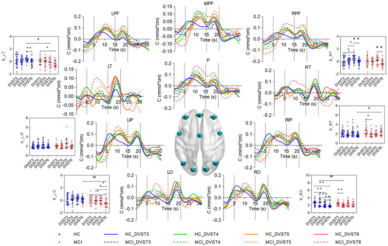
Results: (1) During the encoding phase, both the left occipital (pcond = 0.05, pgroup < 0.01) and left temporal (pcond = 0.02, pgroup = 0.03) skewness condition effects and between-group effects were significant. (2) As the cognitive load increased, the clustering coefficients and local efficiencies were significantly lower for the HC group. (3) The left occipital and left temporal activation skewness in the MCI group were significantly correlated with left occipital electrical features, whereas the left occipital activation intensity and skewness were significantly correlated with left occipital electrical features in HC group.
Conclusions: The pattern of brain activity in MCI depends on cognitive load. Left occipital and left temporal may be important brain regions for evaluating MCI and need to be focused on in the future.

 求助内容:
求助内容: 应助结果提醒方式:


