{"title":"[Anatomy and physiology of the lymph system : What clinicians should know].","authors":"Erich Brenner","doi":"10.1007/s00117-025-01432-2","DOIUrl":null,"url":null,"abstract":"<p><p>The lymphatic vessels are a unidirectional, centripetal system that collects interstitial fluid from tissues and organs, directing it into the venous system. The lymphatic vessels can be categorized into four types: initial lymphatic vessels, precollectors, collectors, and lymphatic trunks. Initial lymphatic vessels comprise interconnected networks characterized by larger mash sizes compared to blood capillaries and are lined with a specialized endothelium. They have interendothelial openings that allow exchange with the surrounding tissue. These vessels also have characteristic markers such as VEGFR‑3, podoplanin, and LYVE‑1. Precollectors have smooth muscle cells that are still arranged irregularly and parietal valves that promote centripetal lymph flow. Collectors have also valves and a continuous layer of spirally arranged muscle fibers that allow the vessels to pulse and contract. This contraction causes the lymph to be transported from one lymphangion to the next. Lymphatic trunks form the next section and often flow into the thoracic duct, which opens into the venous system. Lymph nodes are situated along the course of the collectors and trunks and serve as filter centers to intercept pathogens and increase the colloid osmotic pressure of the lymph. They are organized into groups with a specific structure with cortex, medulla, and embedded B and T lymphocytes. The central lymphatic system transports lymph through segmental connections and anastomoses. The thoracic duct empties into the left venous angle and collects also other lymphatic truncs. The thoracic duct's developmental history and variations occasionally lead to anatomical anomalies.</p>","PeriodicalId":74635,"journal":{"name":"Radiologie (Heidelberg, Germany)","volume":" ","pages":"301-306"},"PeriodicalIF":0.6000,"publicationDate":"2025-05-01","publicationTypes":"Journal Article","fieldsOfStudy":null,"isOpenAccess":false,"openAccessPdf":"https://www.ncbi.nlm.nih.gov/pmc/articles/PMC12040980/pdf/","citationCount":"0","resultStr":null,"platform":"Semanticscholar","paperid":null,"PeriodicalName":"Radiologie (Heidelberg, Germany)","FirstCategoryId":"1085","ListUrlMain":"https://doi.org/10.1007/s00117-025-01432-2","RegionNum":0,"RegionCategory":null,"ArticlePicture":[],"TitleCN":null,"AbstractTextCN":null,"PMCID":null,"EPubDate":"2025/3/22 0:00:00","PubModel":"Epub","JCR":"","JCRName":"","Score":null,"Total":0}
引用次数: 0
Abstract
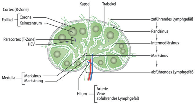
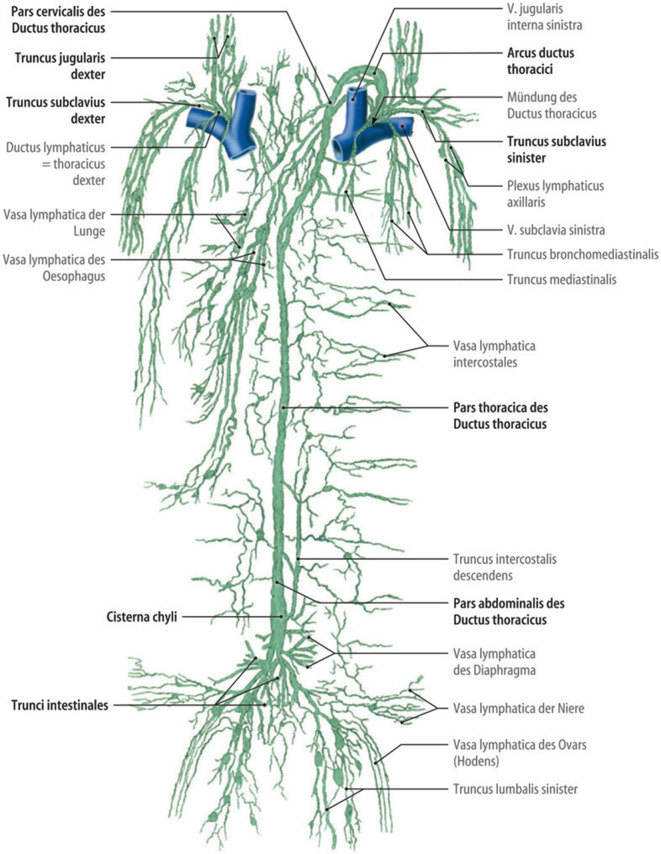
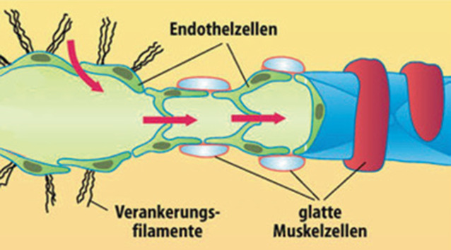
The lymphatic vessels are a unidirectional, centripetal system that collects interstitial fluid from tissues and organs, directing it into the venous system. The lymphatic vessels can be categorized into four types: initial lymphatic vessels, precollectors, collectors, and lymphatic trunks. Initial lymphatic vessels comprise interconnected networks characterized by larger mash sizes compared to blood capillaries and are lined with a specialized endothelium. They have interendothelial openings that allow exchange with the surrounding tissue. These vessels also have characteristic markers such as VEGFR‑3, podoplanin, and LYVE‑1. Precollectors have smooth muscle cells that are still arranged irregularly and parietal valves that promote centripetal lymph flow. Collectors have also valves and a continuous layer of spirally arranged muscle fibers that allow the vessels to pulse and contract. This contraction causes the lymph to be transported from one lymphangion to the next. Lymphatic trunks form the next section and often flow into the thoracic duct, which opens into the venous system. Lymph nodes are situated along the course of the collectors and trunks and serve as filter centers to intercept pathogens and increase the colloid osmotic pressure of the lymph. They are organized into groups with a specific structure with cortex, medulla, and embedded B and T lymphocytes. The central lymphatic system transports lymph through segmental connections and anastomoses. The thoracic duct empties into the left venous angle and collects also other lymphatic truncs. The thoracic duct's developmental history and variations occasionally lead to anatomical anomalies.

 求助内容:
求助内容: 应助结果提醒方式:


