András Molnár, Viktória Molnár, Panayiota Mavrogeni, Stefani Maihoub
{"title":"The Influence of Carotid and Vertebral Doppler Ultrasonography and Brain MRI Abnormalities on Hearing Levels, Tinnitus Intensities and Frequencies.","authors":"András Molnár, Viktória Molnár, Panayiota Mavrogeni, Stefani Maihoub","doi":"10.3390/audiolres15020029","DOIUrl":null,"url":null,"abstract":"<p><p><b>Objectives</b>: This study aimed to analyse the potential influence of abnormalities detected through carotid-vertebral ultrasonography and brain MRI on pure-tone averages (PTAs) and the frequency and intensity of tinnitus. <b>Methods</b>: 423 participants with subjective tinnitus were enrolled in this investigation. All patients underwent carotid- vertebral ultrasonography, brain MRI, and pure-tone audiometry, including tinnitus matching. <b>Results</b>: The median values for tinnitus onset indicated chronic tinnitus in most cases. Regarding tinnitus location, left-sided symptoms (32%) and bilateral symptoms (44%) were the most prevalent. In analysing the effects of abnormalities detected by carotid-vertebral ultrasonography on PTAs, a statistically significant difference was found between the groups (<i>p</i> = 0.0037). Specifically, individuals with intimal hyperplasia had significantly higher PTAs (<i>p</i> = 0.02), as did those with carotid artery plaques (<i>p</i> = 0.005). However, no significant differences in PTAs were noted in relation to carotid artery stenosis (<i>p</i> = 0.07). Similar trends emerged regarding tinnitus intensity (<i>p</i> = 0.013), with significantly higher values observed in the presence of any carotid-vertebral ultrasonography abnormalities. In contrast, tinnitus frequencies were not significantly affected (<i>p</i> = 0.401). Regarding brain MRI findings, Fazekas scores of 2 (<i>p</i> = 0.02) and 3 (<i>p</i> = 0.0052) significantly influenced PTAs. For tinnitus intensity, Fazekas scores of 2 (<i>p</i> = 0.0027) and 3 (<i>p</i> = 0.0005), and the presence of acoustic neuromas <i>(p</i> = 0.019), significantly impacted the intensity values. However, tinnitus frequencies were not significantly (<i>p</i> = 0.36) influenced by brain MRI abnormalities. <b>Conclusions</b>: The findings of this study show that carotid-vertebral ultrasonography and brain MRI abnormalities significantly influence PTAs and tinnitus intensities.</p>","PeriodicalId":44133,"journal":{"name":"Audiology Research","volume":"15 2","pages":""},"PeriodicalIF":1.8000,"publicationDate":"2025-03-15","publicationTypes":"Journal Article","fieldsOfStudy":null,"isOpenAccess":false,"openAccessPdf":"https://www.ncbi.nlm.nih.gov/pmc/articles/PMC11932294/pdf/","citationCount":"0","resultStr":null,"platform":"Semanticscholar","paperid":null,"PeriodicalName":"Audiology Research","FirstCategoryId":"1085","ListUrlMain":"https://doi.org/10.3390/audiolres15020029","RegionNum":0,"RegionCategory":null,"ArticlePicture":[],"TitleCN":null,"AbstractTextCN":null,"PMCID":null,"EPubDate":"","PubModel":"","JCR":"Q1","JCRName":"AUDIOLOGY & SPEECH-LANGUAGE PATHOLOGY","Score":null,"Total":0}
引用次数: 0
Abstract
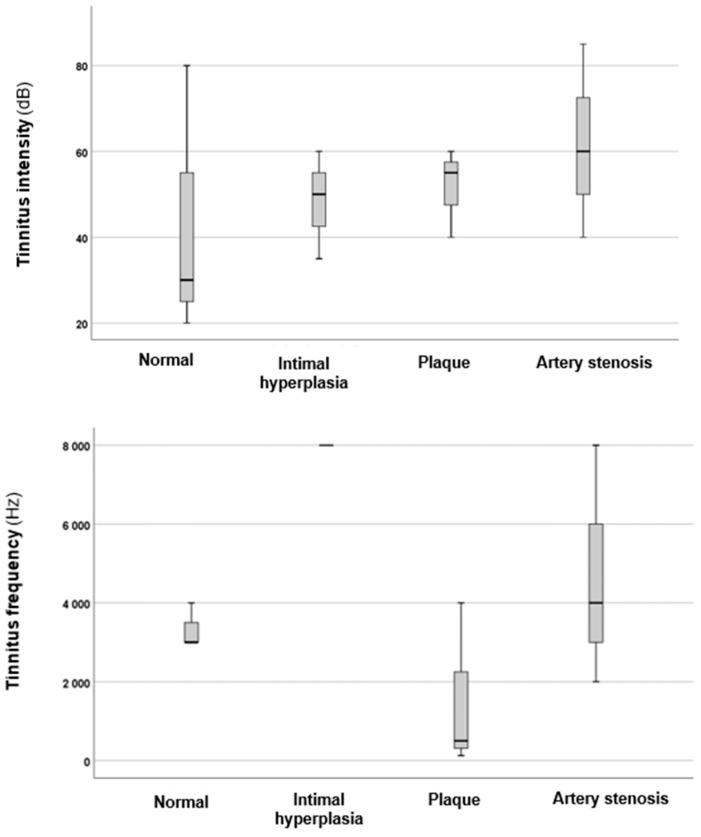
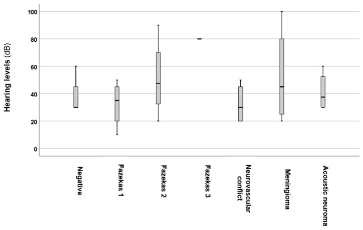
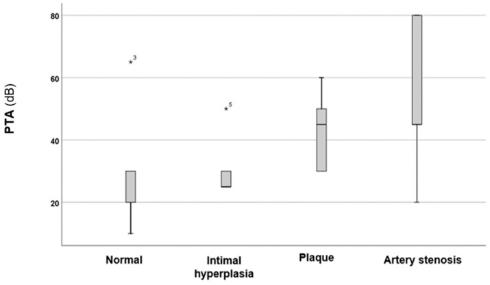
Objectives: This study aimed to analyse the potential influence of abnormalities detected through carotid-vertebral ultrasonography and brain MRI on pure-tone averages (PTAs) and the frequency and intensity of tinnitus. Methods: 423 participants with subjective tinnitus were enrolled in this investigation. All patients underwent carotid- vertebral ultrasonography, brain MRI, and pure-tone audiometry, including tinnitus matching. Results: The median values for tinnitus onset indicated chronic tinnitus in most cases. Regarding tinnitus location, left-sided symptoms (32%) and bilateral symptoms (44%) were the most prevalent. In analysing the effects of abnormalities detected by carotid-vertebral ultrasonography on PTAs, a statistically significant difference was found between the groups (p = 0.0037). Specifically, individuals with intimal hyperplasia had significantly higher PTAs (p = 0.02), as did those with carotid artery plaques (p = 0.005). However, no significant differences in PTAs were noted in relation to carotid artery stenosis (p = 0.07). Similar trends emerged regarding tinnitus intensity (p = 0.013), with significantly higher values observed in the presence of any carotid-vertebral ultrasonography abnormalities. In contrast, tinnitus frequencies were not significantly affected (p = 0.401). Regarding brain MRI findings, Fazekas scores of 2 (p = 0.02) and 3 (p = 0.0052) significantly influenced PTAs. For tinnitus intensity, Fazekas scores of 2 (p = 0.0027) and 3 (p = 0.0005), and the presence of acoustic neuromas (p = 0.019), significantly impacted the intensity values. However, tinnitus frequencies were not significantly (p = 0.36) influenced by brain MRI abnormalities. Conclusions: The findings of this study show that carotid-vertebral ultrasonography and brain MRI abnormalities significantly influence PTAs and tinnitus intensities.
期刊介绍:
The mission of Audiology Research is to publish contemporary, ethical, clinically relevant scientific researches related to the basic science and clinical aspects of the auditory and vestibular system and diseases of the ear that can be used by clinicians, scientists and specialists to improve understanding and treatment of patients with audiological and neurotological disorders.

 求助内容:
求助内容: 应助结果提醒方式:


