{"title":"Optical coherence tomography findings in patients with diabetic macular edema: A retrospective analysis.","authors":"Manjunathan Sivarasu, Gopinath Madheswaran, Saranya Sachi Balasubramaniam, Chinnasamy Balasubramaniam","doi":"10.4103/ojo.ojo_23_24","DOIUrl":null,"url":null,"abstract":"<p><strong>Background: </strong>Diabetic macular edema (DME) is a leading cause of vision loss in diabetic people. DME can be treated with various medications, including intravitreal injections, laser therapy, and surgery. Early detection and treatment of DME is essential to prevent vision loss. The study aimed to describe patients' demographic and clinical characteristics with DME, optical coherence tomography (OCT) findings, and visual acuity outcomes.</p><p><strong>Methodology: </strong>A retrospective study reviewed case records of patients with DME between 2017 and 2020. Demographic data, clinical characteristics, and examination results were extracted and analyzed using Microsoft Excel (2013). All patients clinically diagnosed with DME underwent assessment by OCT examination. DME was classified based on OCT findings. Statistical significance was observed at <i>P</i> < 0.05.</p><p><strong>Results: </strong>This retrospective study included 213 eyes of 134 patients, of which 77.6% were male and 22.4% were female. Nonproliferative diabetic retinopathy (NPDR) was present in 51.64% of eyes, and PDR was present in 48.36%. Focal, diffuse, and cystoid macular edema was observed in 68, 31, and 65 eyes, respectively. Tractional macular edema was seen in 16 eyes with posterior hyaloid traction, 13 with epiretinal membrane (ERM), and one with both conditions. DME associated with subretinal fluid (SRF) detachment was seen in 8.92% of eyes. The mean (standard deviation) central retinal thickness was 284.5 (28.9), 434.0 (97.5), 426.5 (27.5), 510.5 (14.1), and 465.5 (280.7) μm in focal, diffuse, cystoid, ERM, and SRF, respectively. Increased central retinal thickness was associated with decreased visual acuity (<i>P</i> < 0.05).</p><p><strong>Conclusion: </strong>The findings of this study suggest that DME is a common and visually significant complication of diabetes. The OCT findings can be used to classify DME into different subtypes, which may help to guide treatment decisions. Focal edema was the most common type of DME with the least central retinal thickness. In NPDR, focal macular edema was the most common; in PDR, cystoid edema was the most common. Cystoid edema was the most common type in the subgroup of patients with recurrent DME following anti-vascular endothelial growth factor injection.</p>","PeriodicalId":19461,"journal":{"name":"Oman Journal of Ophthalmology","volume":"18 1","pages":"22-27"},"PeriodicalIF":0.0000,"publicationDate":"2025-02-25","publicationTypes":"Journal Article","fieldsOfStudy":null,"isOpenAccess":false,"openAccessPdf":"https://www.ncbi.nlm.nih.gov/pmc/articles/PMC11925365/pdf/","citationCount":"0","resultStr":null,"platform":"Semanticscholar","paperid":null,"PeriodicalName":"Oman Journal of Ophthalmology","FirstCategoryId":"1085","ListUrlMain":"https://doi.org/10.4103/ojo.ojo_23_24","RegionNum":0,"RegionCategory":null,"ArticlePicture":[],"TitleCN":null,"AbstractTextCN":null,"PMCID":null,"EPubDate":"2025/1/1 0:00:00","PubModel":"eCollection","JCR":"Q3","JCRName":"Medicine","Score":null,"Total":0}
引用次数: 0
Abstract
Background: Diabetic macular edema (DME) is a leading cause of vision loss in diabetic people. DME can be treated with various medications, including intravitreal injections, laser therapy, and surgery. Early detection and treatment of DME is essential to prevent vision loss. The study aimed to describe patients' demographic and clinical characteristics with DME, optical coherence tomography (OCT) findings, and visual acuity outcomes.
Methodology: A retrospective study reviewed case records of patients with DME between 2017 and 2020. Demographic data, clinical characteristics, and examination results were extracted and analyzed using Microsoft Excel (2013). All patients clinically diagnosed with DME underwent assessment by OCT examination. DME was classified based on OCT findings. Statistical significance was observed at P < 0.05.
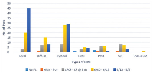
Results: This retrospective study included 213 eyes of 134 patients, of which 77.6% were male and 22.4% were female. Nonproliferative diabetic retinopathy (NPDR) was present in 51.64% of eyes, and PDR was present in 48.36%. Focal, diffuse, and cystoid macular edema was observed in 68, 31, and 65 eyes, respectively. Tractional macular edema was seen in 16 eyes with posterior hyaloid traction, 13 with epiretinal membrane (ERM), and one with both conditions. DME associated with subretinal fluid (SRF) detachment was seen in 8.92% of eyes. The mean (standard deviation) central retinal thickness was 284.5 (28.9), 434.0 (97.5), 426.5 (27.5), 510.5 (14.1), and 465.5 (280.7) μm in focal, diffuse, cystoid, ERM, and SRF, respectively. Increased central retinal thickness was associated with decreased visual acuity (P < 0.05).
Conclusion: The findings of this study suggest that DME is a common and visually significant complication of diabetes. The OCT findings can be used to classify DME into different subtypes, which may help to guide treatment decisions. Focal edema was the most common type of DME with the least central retinal thickness. In NPDR, focal macular edema was the most common; in PDR, cystoid edema was the most common. Cystoid edema was the most common type in the subgroup of patients with recurrent DME following anti-vascular endothelial growth factor injection.
期刊介绍:
To provide a platform for scientific expression of the Oman Ophthalmic Society and the international Ophthalmic community and to provide opportunities for free exchange of ideas and information. To serve as a valuable resource for ophthalmologists, eye-care providers including optometrists, orthoptists, other health care professionals and research workers in all aspects of the field of visual science.

 求助内容:
求助内容: 应助结果提醒方式:


