{"title":"Scale Surface Topography of a Vulnerable Cyprinid Fish, Schizothorax plagiostomus From Kashmir Himalayas Using Scanning Electron Microscopy (SEM)","authors":"Misba Rehman, Syed Talia Mushtaq, Tasaduq Hussain Shah, Farooz Ahmad Bhat","doi":"10.1002/jemt.24861","DOIUrl":null,"url":null,"abstract":"<div>\n \n <p>Scanning electron microscopy (SEM) has significantly advanced morphological studies, particularly in the investigation of fish scale structures. This technique has unveiled intricate architectural details that are crucial for fish identification and classification. In this study, macro and microscopic analyses were employed to examine the scale morphology of <i>Schizothorax plagiostomus</i>, a vulnerable cyprinid fish from Kashmir, focusing on two body regions (region below dorsal fin and above lateral line). The general scale type observed in <i>S. plagiostomus</i> was cycloid. Two types of shapes, viz., polygonal and cordate, were reported in this species. The rostral margin of the scales displayed round and waved forms. The scales exhibited a small and round focus, which was antero-centrally positioned. The scales featured narrow or wide grooves (radii) categorized into three types: primary, secondary, and tertiary, present across all four scale fields (anterior, posterior, dorsal and lateral), forming a tetra-sectioned type. Circuli, arranged in circular patterns around the focus, were present, which were densely placed in the anterior and lateral fields and widely spaced in the posterior field. Notably, lepidonts on the circuli and chromatophores on the posterior margin were absent in this species. These scale characteristics and their morphologies offer a valuable tool for the identification, classification, and phylogenetic analysis of various freshwater fish species and genera.</p>\n </div>","PeriodicalId":18684,"journal":{"name":"Microscopy Research and Technique","volume":"88 8","pages":"2310-2320"},"PeriodicalIF":2.1000,"publicationDate":"2025-03-23","publicationTypes":"Journal Article","fieldsOfStudy":null,"isOpenAccess":false,"openAccessPdf":"","citationCount":"0","resultStr":null,"platform":"Semanticscholar","paperid":null,"PeriodicalName":"Microscopy Research and Technique","FirstCategoryId":"5","ListUrlMain":"https://analyticalsciencejournals.onlinelibrary.wiley.com/doi/10.1002/jemt.24861","RegionNum":3,"RegionCategory":"工程技术","ArticlePicture":[],"TitleCN":null,"AbstractTextCN":null,"PMCID":null,"EPubDate":"","PubModel":"","JCR":"Q2","JCRName":"ANATOMY & MORPHOLOGY","Score":null,"Total":0}
引用次数: 0
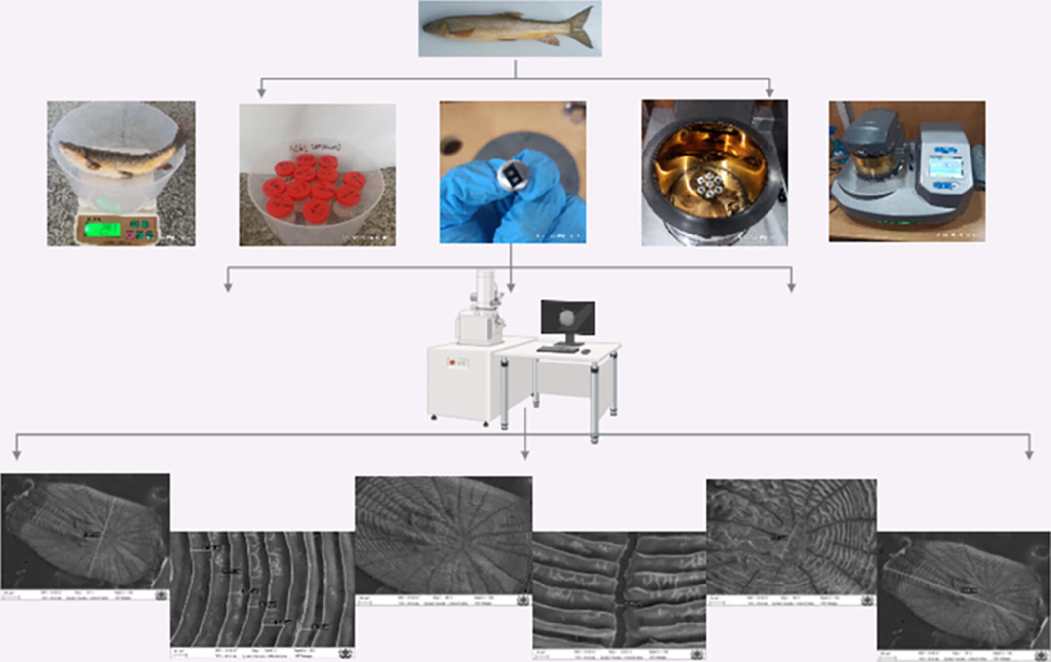
Abstract
Scanning electron microscopy (SEM) has significantly advanced morphological studies, particularly in the investigation of fish scale structures. This technique has unveiled intricate architectural details that are crucial for fish identification and classification. In this study, macro and microscopic analyses were employed to examine the scale morphology of Schizothorax plagiostomus, a vulnerable cyprinid fish from Kashmir, focusing on two body regions (region below dorsal fin and above lateral line). The general scale type observed in S. plagiostomus was cycloid. Two types of shapes, viz., polygonal and cordate, were reported in this species. The rostral margin of the scales displayed round and waved forms. The scales exhibited a small and round focus, which was antero-centrally positioned. The scales featured narrow or wide grooves (radii) categorized into three types: primary, secondary, and tertiary, present across all four scale fields (anterior, posterior, dorsal and lateral), forming a tetra-sectioned type. Circuli, arranged in circular patterns around the focus, were present, which were densely placed in the anterior and lateral fields and widely spaced in the posterior field. Notably, lepidonts on the circuli and chromatophores on the posterior margin were absent in this species. These scale characteristics and their morphologies offer a valuable tool for the identification, classification, and phylogenetic analysis of various freshwater fish species and genera.
期刊介绍:
Microscopy Research and Technique (MRT) publishes articles on all aspects of advanced microscopy original architecture and methodologies with applications in the biological, clinical, chemical, and materials sciences. Original basic and applied research as well as technical papers dealing with the various subsets of microscopy are encouraged. MRT is the right form for those developing new microscopy methods or using the microscope to answer key questions in basic and applied research.


 求助内容:
求助内容: 应助结果提醒方式:


