{"title":"A Case of Omental Neuroendocrine Tumor Discovered Incidentally: Case Report.","authors":"Masataka Taki, Toshikatsu Nitta, Ryutaro Kubo, Aki Yoshiyama, Hidero Yoshimoto, Masatsugu Ishii, Takashi Ishibashi, Atsushi Takeshita","doi":"10.1177/23247096241299286","DOIUrl":null,"url":null,"abstract":"<p><p>Neuroendocrine cells are distributed throughout the body's organs, though neuroendocrine neoplasms are primarily documented in the gastrointestinal tract and pancreas, with rare occurrences elsewhere. Herein, we report a case of primary neuroendocrine tumor of the omentum (omental NET) that was incidentally detected as an omental mass during preoperative screening for colorectal cancer. The patient, a 66-year-old woman, with abdominal pain and decreased oral intake, leading to a diagnosis of obstructive colorectal cancer with a large, 55 mm, mass around the gastropyloric region, which was discontinuous with the gastrointestinal tract. After the placement of a colonic stent at the site of the ascending colon cancer to decompress the colon, a laparoscopic right hemicolectomy was performed, simultaneously excising the mass. Postoperative pathology revealed a neuroendocrine tumor (NET). Subsequent examinations detected no other lesions of suspected primary disease and postoperative somatostatin scintigraphy found no other lesions, establishing a diagnosis of omental NET. The rarity of omental NETs is attributable to the absence of neuroendocrine cells in the omentum. Moreover, solid tumors originating primarily from the omentum are very rare, making preoperative diagnosis difficult; therefore, postoperative pathology should be utilized. We presented a very rare case of omental NET, previously reported only once in the literature, and believe that complete resection with minimal invasiveness should be performed for treatment of this malignancy. In addition, we emphasize the need for continued patient follow-up.</p>","PeriodicalId":16198,"journal":{"name":"Journal of investigative medicine high impact case reports","volume":"13 ","pages":"23247096241299286"},"PeriodicalIF":0.8000,"publicationDate":"2025-01-01","publicationTypes":"Journal Article","fieldsOfStudy":null,"isOpenAccess":false,"openAccessPdf":"https://www.ncbi.nlm.nih.gov/pmc/articles/PMC11938437/pdf/","citationCount":"0","resultStr":null,"platform":"Semanticscholar","paperid":null,"PeriodicalName":"Journal of investigative medicine high impact case reports","FirstCategoryId":"1085","ListUrlMain":"https://doi.org/10.1177/23247096241299286","RegionNum":0,"RegionCategory":null,"ArticlePicture":[],"TitleCN":null,"AbstractTextCN":null,"PMCID":null,"EPubDate":"2025/3/24 0:00:00","PubModel":"Epub","JCR":"Q3","JCRName":"MEDICINE, GENERAL & INTERNAL","Score":null,"Total":0}
引用次数: 0
Abstract
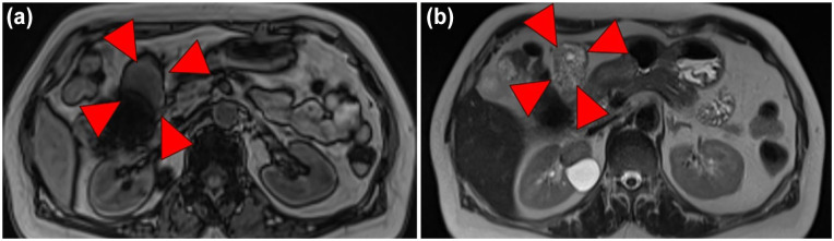
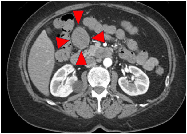
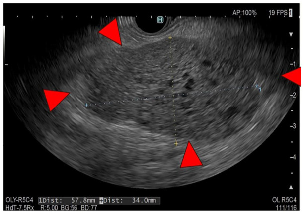
Neuroendocrine cells are distributed throughout the body's organs, though neuroendocrine neoplasms are primarily documented in the gastrointestinal tract and pancreas, with rare occurrences elsewhere. Herein, we report a case of primary neuroendocrine tumor of the omentum (omental NET) that was incidentally detected as an omental mass during preoperative screening for colorectal cancer. The patient, a 66-year-old woman, with abdominal pain and decreased oral intake, leading to a diagnosis of obstructive colorectal cancer with a large, 55 mm, mass around the gastropyloric region, which was discontinuous with the gastrointestinal tract. After the placement of a colonic stent at the site of the ascending colon cancer to decompress the colon, a laparoscopic right hemicolectomy was performed, simultaneously excising the mass. Postoperative pathology revealed a neuroendocrine tumor (NET). Subsequent examinations detected no other lesions of suspected primary disease and postoperative somatostatin scintigraphy found no other lesions, establishing a diagnosis of omental NET. The rarity of omental NETs is attributable to the absence of neuroendocrine cells in the omentum. Moreover, solid tumors originating primarily from the omentum are very rare, making preoperative diagnosis difficult; therefore, postoperative pathology should be utilized. We presented a very rare case of omental NET, previously reported only once in the literature, and believe that complete resection with minimal invasiveness should be performed for treatment of this malignancy. In addition, we emphasize the need for continued patient follow-up.
期刊介绍:
The AFMR is committed to enhancing the training and career development of our members and to furthering its mission to facilitate the conduct of research to improve medical care. Case reports represent an important avenue for trainees (interns, residents, and fellows) and early-stage faculty to demonstrate productive, scholarly activity.

 求助内容:
求助内容: 应助结果提醒方式:


