Monica Cheng, Nikita Consul, Ryan Chung, Carlos Fernandez- Del Castillo, Yasmin Hernandez-Barco, Avinash Kambadakone
{"title":"Acinar cell carcinoma of the pancreas: can CT and MR features predict survival?","authors":"Monica Cheng, Nikita Consul, Ryan Chung, Carlos Fernandez- Del Castillo, Yasmin Hernandez-Barco, Avinash Kambadakone","doi":"10.1186/s40644-025-00859-z","DOIUrl":null,"url":null,"abstract":"<p><strong>Objective: </strong>To evaluate the CT and MRI features of pancreatic acinar cell carcinoma (pACC) and their association with patient outcome and survival.</p><p><strong>Methods: </strong>This retrospective single-center study included 49 patients with pathology-proven pancreatic acinar cell carcinoma who underwent diagnostic imaging between August 1998 - September 2019. Two radiologists reviewed CT and MRI features independently. Survival was estimated using the Kaplan-Meier method, and Cox proportional-hazards regression model was used to identify factors associated with survival.</p><p><strong>Results: </strong>pACC tended to present as a solid (31/49, 63.3%) pancreatic head mass (26/49, 53.1%) with ill-defined margins (32/49, 65.3%) and median maximal diameter of 4.1 cm (IQR, 2.9-6.2). Majority of lesions were hypo- or isodense (38/49, 77.6%) compared to normal pancreatic parenchyma, with heterogenous (39/49, 79.6%) enhancement pattern. Biliary ductal dilatation was uncommon, with pancreatic ductal dilatation in 22.4% (11/49) and common bile duct dilatation in 14.3% (7/49). Intralesional calcifications were seen in 6.1% (3/49). Metastasis was present in 71.4% (35/49) of patients at the time of diagnosis. On MRI, 88.9% (16/18) demonstrated diffusion restriction and 59.1% (13/22) with heterogenous enhancement. On multivariate analysis, the imaging presence of T1 hyperintensity (p = 0.02), hypoattenuating necrotic components (p = 0.02), and splenic vein invasion (p = 0.04) were associated with worse survival.</p><p><strong>Conclusion: </strong>Pancreatic acinar cell carcinoma is a rare pancreatic neoplasm that often presents as a large ill-defined heterogeneously enhancing mass without biliary ductal dilation. T1 hyperintensity, presence of hypoattenuating necrotic components, and splenic vein invasion were independent predictors of survival.</p>","PeriodicalId":9548,"journal":{"name":"Cancer Imaging","volume":"25 1","pages":"38"},"PeriodicalIF":3.5000,"publicationDate":"2025-03-21","publicationTypes":"Journal Article","fieldsOfStudy":null,"isOpenAccess":false,"openAccessPdf":"https://www.ncbi.nlm.nih.gov/pmc/articles/PMC11929164/pdf/","citationCount":"0","resultStr":null,"platform":"Semanticscholar","paperid":null,"PeriodicalName":"Cancer Imaging","FirstCategoryId":"3","ListUrlMain":"https://doi.org/10.1186/s40644-025-00859-z","RegionNum":2,"RegionCategory":"医学","ArticlePicture":[],"TitleCN":null,"AbstractTextCN":null,"PMCID":null,"EPubDate":"","PubModel":"","JCR":"Q2","JCRName":"ONCOLOGY","Score":null,"Total":0}
引用次数: 0
Abstract
Objective: To evaluate the CT and MRI features of pancreatic acinar cell carcinoma (pACC) and their association with patient outcome and survival.
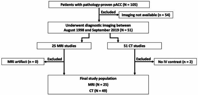
Methods: This retrospective single-center study included 49 patients with pathology-proven pancreatic acinar cell carcinoma who underwent diagnostic imaging between August 1998 - September 2019. Two radiologists reviewed CT and MRI features independently. Survival was estimated using the Kaplan-Meier method, and Cox proportional-hazards regression model was used to identify factors associated with survival.
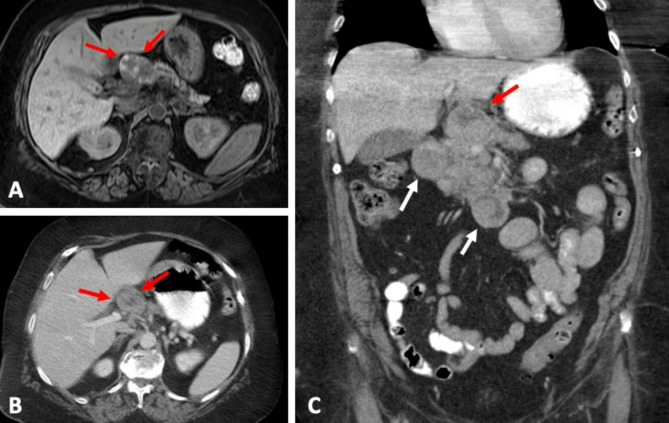
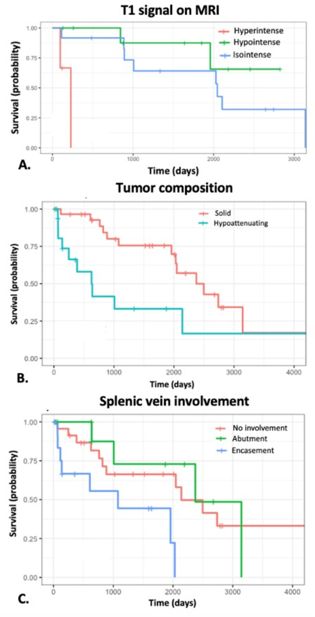
Results: pACC tended to present as a solid (31/49, 63.3%) pancreatic head mass (26/49, 53.1%) with ill-defined margins (32/49, 65.3%) and median maximal diameter of 4.1 cm (IQR, 2.9-6.2). Majority of lesions were hypo- or isodense (38/49, 77.6%) compared to normal pancreatic parenchyma, with heterogenous (39/49, 79.6%) enhancement pattern. Biliary ductal dilatation was uncommon, with pancreatic ductal dilatation in 22.4% (11/49) and common bile duct dilatation in 14.3% (7/49). Intralesional calcifications were seen in 6.1% (3/49). Metastasis was present in 71.4% (35/49) of patients at the time of diagnosis. On MRI, 88.9% (16/18) demonstrated diffusion restriction and 59.1% (13/22) with heterogenous enhancement. On multivariate analysis, the imaging presence of T1 hyperintensity (p = 0.02), hypoattenuating necrotic components (p = 0.02), and splenic vein invasion (p = 0.04) were associated with worse survival.
Conclusion: Pancreatic acinar cell carcinoma is a rare pancreatic neoplasm that often presents as a large ill-defined heterogeneously enhancing mass without biliary ductal dilation. T1 hyperintensity, presence of hypoattenuating necrotic components, and splenic vein invasion were independent predictors of survival.
Cancer ImagingONCOLOGY-RADIOLOGY, NUCLEAR MEDICINE & MEDICAL IMAGING
CiteScore
7.00
自引率
0.00%
发文量
66
审稿时长
>12 weeks
期刊介绍:
Cancer Imaging is an open access, peer-reviewed journal publishing original articles, reviews and editorials written by expert international radiologists working in oncology.
The journal encompasses CT, MR, PET, ultrasound, radionuclide and multimodal imaging in all kinds of malignant tumours, plus new developments, techniques and innovations. Topics of interest include:
Breast Imaging
Chest
Complications of treatment
Ear, Nose & Throat
Gastrointestinal
Hepatobiliary & Pancreatic
Imaging biomarkers
Interventional
Lymphoma
Measurement of tumour response
Molecular functional imaging
Musculoskeletal
Neuro oncology
Nuclear Medicine
Paediatric.

 求助内容:
求助内容: 应助结果提醒方式:


