Ana Paula Vargas Garcia, Fernanda Rezende Souza, Luiz Flávio Telles, Antônio Carlos Lacreta, Mary Suzan Varaschin, Geovanni Dantas Cassali
{"title":"Histopathological, immunohistochemical and imaging features of bone metastases of mammary carcinoma in bitches: cases report.","authors":"Ana Paula Vargas Garcia, Fernanda Rezende Souza, Luiz Flávio Telles, Antônio Carlos Lacreta, Mary Suzan Varaschin, Geovanni Dantas Cassali","doi":"10.29374/2527-2179.bjvm009124","DOIUrl":null,"url":null,"abstract":"<p><p>In the present case report, the histopathological and immunohistochemical characteristics of the two cases of bone metastasis of mammary carcinoma in bitches are described. The animal in the first case is a 10 years old female poodle. The physical examination revealed a mass in the left abdominal caudal (M4) and inguinal (M5) mammary glands with a six-month evolution. The imaging exams of the right pelvic limb revealed areas of bone lysis in the distal portion of the femur. No evidence of metastases was observed in the thorax on thoracic radiographs. Microscopic evaluations were consistent with the diagnosis of malignant adenomyoepithelioma. The mass in the distal region of the femur has characteristics similar to those observed in the mammary gland mass. The animal in the second case was a nine-year-old female mixed-breed euthanized due to the unfavorable prognosis of the disease. Histopathological evaluation of the primary tumor in M3, M4, and M5 was consistent with the diagnosis of grade II cribriform carcinoma. Metastatic foci were observed in the lung, liver, kidney, adrenal, proximal metaphyseal region of the right humerus extending to the distal diaphyseal region, and axillary and medial iliac lymph nodes' parenchyma. Immunohistochemistry was performed for markers Ki67, Cox-2, ER, PR, Pan-CK, p63 and HER-2 in the primary tumor and bone metastasis in both cases. High proliferation rate, positivity for hormone receptors, Pan-CK and p63 were observed in both cases. HER-2 was negative in the primary tumor and bone metastasis and COX-2 was negative in the primary tumor of both cases, negative in the metastasis of case 01 and positive in the metastasis of case 02.</p>","PeriodicalId":72458,"journal":{"name":"Brazilian journal of veterinary medicine","volume":"47 ","pages":"e009124"},"PeriodicalIF":0.0000,"publicationDate":"2025-03-19","publicationTypes":"Journal Article","fieldsOfStudy":null,"isOpenAccess":false,"openAccessPdf":"https://www.ncbi.nlm.nih.gov/pmc/articles/PMC11924272/pdf/","citationCount":"0","resultStr":null,"platform":"Semanticscholar","paperid":null,"PeriodicalName":"Brazilian journal of veterinary medicine","FirstCategoryId":"1085","ListUrlMain":"https://doi.org/10.29374/2527-2179.bjvm009124","RegionNum":0,"RegionCategory":null,"ArticlePicture":[],"TitleCN":null,"AbstractTextCN":null,"PMCID":null,"EPubDate":"2025/1/1 0:00:00","PubModel":"eCollection","JCR":"","JCRName":"","Score":null,"Total":0}
引用次数: 0
Abstract
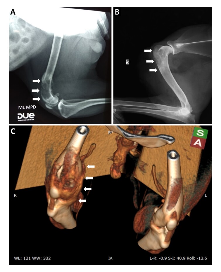
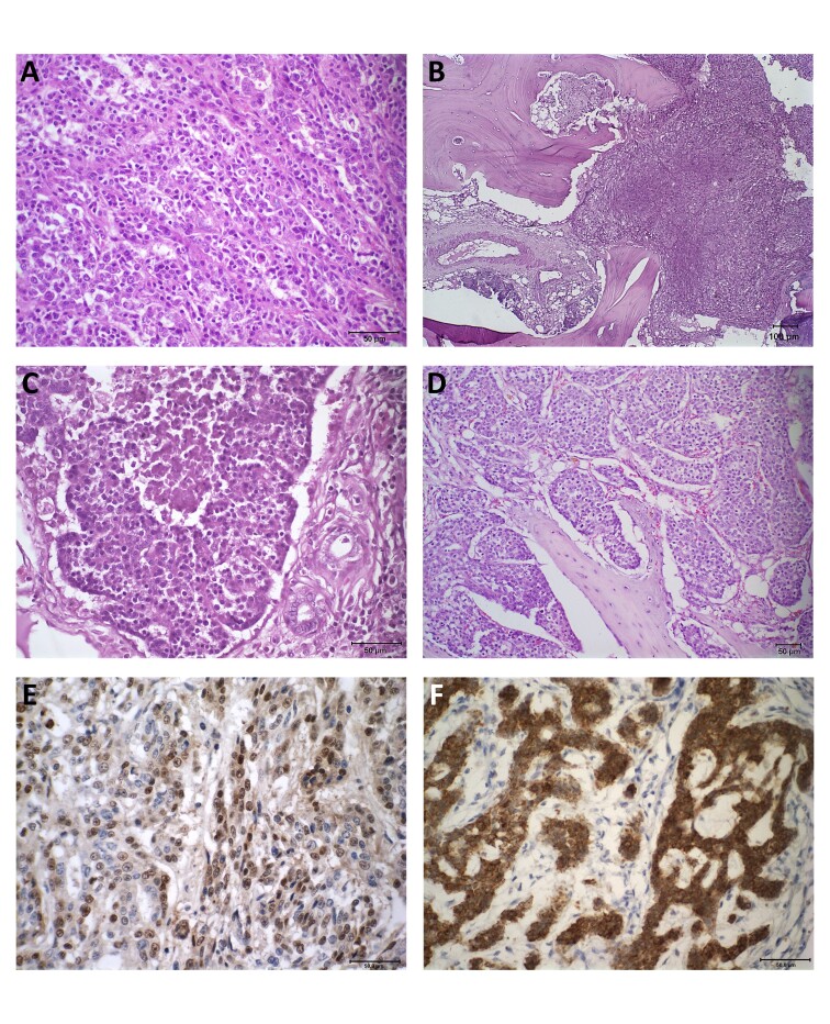
In the present case report, the histopathological and immunohistochemical characteristics of the two cases of bone metastasis of mammary carcinoma in bitches are described. The animal in the first case is a 10 years old female poodle. The physical examination revealed a mass in the left abdominal caudal (M4) and inguinal (M5) mammary glands with a six-month evolution. The imaging exams of the right pelvic limb revealed areas of bone lysis in the distal portion of the femur. No evidence of metastases was observed in the thorax on thoracic radiographs. Microscopic evaluations were consistent with the diagnosis of malignant adenomyoepithelioma. The mass in the distal region of the femur has characteristics similar to those observed in the mammary gland mass. The animal in the second case was a nine-year-old female mixed-breed euthanized due to the unfavorable prognosis of the disease. Histopathological evaluation of the primary tumor in M3, M4, and M5 was consistent with the diagnosis of grade II cribriform carcinoma. Metastatic foci were observed in the lung, liver, kidney, adrenal, proximal metaphyseal region of the right humerus extending to the distal diaphyseal region, and axillary and medial iliac lymph nodes' parenchyma. Immunohistochemistry was performed for markers Ki67, Cox-2, ER, PR, Pan-CK, p63 and HER-2 in the primary tumor and bone metastasis in both cases. High proliferation rate, positivity for hormone receptors, Pan-CK and p63 were observed in both cases. HER-2 was negative in the primary tumor and bone metastasis and COX-2 was negative in the primary tumor of both cases, negative in the metastasis of case 01 and positive in the metastasis of case 02.

 求助内容:
求助内容: 应助结果提醒方式:


