Avraham Adelman, Landon Richardson, Nikita Chapurin, Brian C Lobo, Si Chen
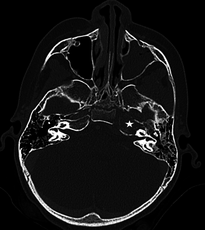
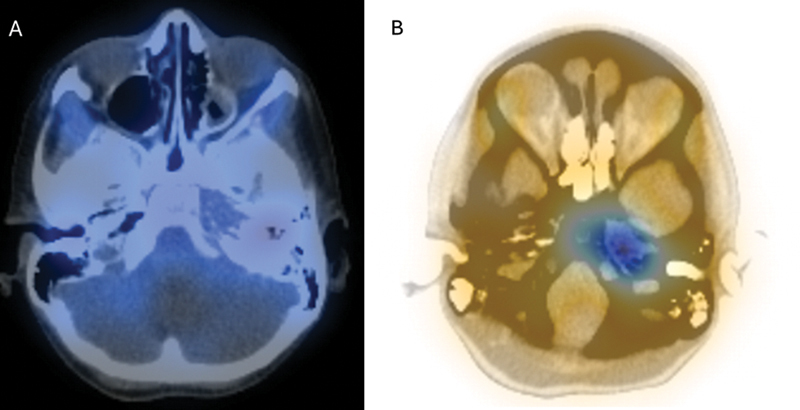
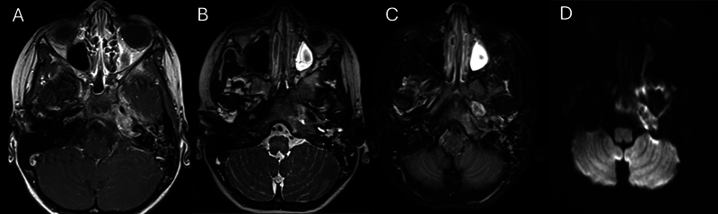
{"title":"Skull Base Rhabdomyosarcoma Mimicking Osteomyelitis in a Pediatric Patient.","authors":"Avraham Adelman, Landon Richardson, Nikita Chapurin, Brian C Lobo, Si Chen","doi":"10.1055/a-2544-3543","DOIUrl":null,"url":null,"abstract":"<p><p>Rhabdomyosarcoma (RMS) is a rare malignant tumor, affecting 4.58 per 1 million children, with approximately 35% occurring in the head and neck. Skull base RMS commonly presents at advanced stages and delays diagnosis due to its overlapping features with other skull base pathology, and difficulty accessing the lesion for biopsy. This case illustrates these challenges in skull base RMS mimicking osteomyelitis of the petrous apex. Case: A 6-year-old immunocompetent female, with a history of two acute otitis media episodes, presented with a 3-week history of sixth cranial nerve palsy and sudden-onset complete seventh cranial nerve palsy. She did not have pain or otorrhea. Computed tomography (CT) and magnetic resonance imaging revealed a 1.3 cm left petrous apex enhancing lesion with extension into the mastoid and clivus with surrounding bony and soft tissue destruction. A nuclear medicine scan (Technetium-99m followed by gallium) demonstrated avid uptake in the left petrous apex. The working diagnosis was skull base osteomyelitis, for which the patient received 2.5 weeks of antibiotics. After failing to improve, repeat imaging showed significant progression of the disease and extension into the nasopharynx and sphenoid sinus. An endoscopic trans-sphenoidal biopsy was performed with pathology consistent with RMS. CT chest revealed lung metastases. The patient partially responded to chemotherapy with vincristine, actinomycin-D, and cyclophosphamide alternating with vincristine and irinotecan. During week 13 of chemotherapy, she received concomitant proton therapy to a total dose of 5040 cGyRBE. Five months after diagnosis, she developed leptomeningeal spread, which was further complicated by meningitis, and passed away.</p>","PeriodicalId":44256,"journal":{"name":"Journal of Neurological Surgery Reports","volume":"86 1","pages":"e41-e44"},"PeriodicalIF":0.7000,"publicationDate":"2025-03-20","publicationTypes":"Journal Article","fieldsOfStudy":null,"isOpenAccess":false,"openAccessPdf":"https://www.ncbi.nlm.nih.gov/pmc/articles/PMC11925613/pdf/","citationCount":"0","resultStr":null,"platform":"Semanticscholar","paperid":null,"PeriodicalName":"Journal of Neurological Surgery Reports","FirstCategoryId":"1085","ListUrlMain":"https://doi.org/10.1055/a-2544-3543","RegionNum":0,"RegionCategory":null,"ArticlePicture":[],"TitleCN":null,"AbstractTextCN":null,"PMCID":null,"EPubDate":"2025/1/1 0:00:00","PubModel":"eCollection","JCR":"Q4","JCRName":"CLINICAL NEUROLOGY","Score":null,"Total":0}
引用次数: 0
Abstract
Rhabdomyosarcoma (RMS) is a rare malignant tumor, affecting 4.58 per 1 million children, with approximately 35% occurring in the head and neck. Skull base RMS commonly presents at advanced stages and delays diagnosis due to its overlapping features with other skull base pathology, and difficulty accessing the lesion for biopsy. This case illustrates these challenges in skull base RMS mimicking osteomyelitis of the petrous apex. Case: A 6-year-old immunocompetent female, with a history of two acute otitis media episodes, presented with a 3-week history of sixth cranial nerve palsy and sudden-onset complete seventh cranial nerve palsy. She did not have pain or otorrhea. Computed tomography (CT) and magnetic resonance imaging revealed a 1.3 cm left petrous apex enhancing lesion with extension into the mastoid and clivus with surrounding bony and soft tissue destruction. A nuclear medicine scan (Technetium-99m followed by gallium) demonstrated avid uptake in the left petrous apex. The working diagnosis was skull base osteomyelitis, for which the patient received 2.5 weeks of antibiotics. After failing to improve, repeat imaging showed significant progression of the disease and extension into the nasopharynx and sphenoid sinus. An endoscopic trans-sphenoidal biopsy was performed with pathology consistent with RMS. CT chest revealed lung metastases. The patient partially responded to chemotherapy with vincristine, actinomycin-D, and cyclophosphamide alternating with vincristine and irinotecan. During week 13 of chemotherapy, she received concomitant proton therapy to a total dose of 5040 cGyRBE. Five months after diagnosis, she developed leptomeningeal spread, which was further complicated by meningitis, and passed away.

 求助内容:
求助内容: 应助结果提醒方式:


