{"title":"Esophageal Atresia Caused by Corrosive Esophagitis for over 50 Years: A Case Report.","authors":"Keisuke Fujimoto, Seiya Inoue, Masakazu Goto, Shinichi Sakamoto, Mariko Misaki, Satoshi Fujiwara, Takahiro Yoshida, Hiroaki Toba, Hiromitsu Takizawa","doi":"10.70352/scrj.cr.24-0116","DOIUrl":null,"url":null,"abstract":"<p><strong>Introduction: </strong>Corrosive esophagitis, often caused by the ingestion of alkalis, acids, or heavy metals, can result in severe esophageal damage and complications, such as stenosis or closure. Although initial treatment is conservative, surgical intervention is necessary when a chronic stricture occurs. A case of esophageal atresia persisting for 50 years due to corrosive esophagitis has not yet been reported. Here, we describe such a case.</p><p><strong>Case presentation: </strong>The patient was a 72-year-old woman. At 20 years of age, she ingested an alkali substance in a suicide attempt, leading to the development of corrosive esophagitis. Surgery was initially considered for esophageal atresia but was deemed unfeasible at the time; therefore, gastrostomy was performed instead. Subsequently, for over 50 years, she manually chewed food and inserted it into her gastric tube. She was urgently transported to a nearby hospital after her general condition deteriorated due to an influenza infection. During hospitalization, her nutritional intake was reassessed, and given her strong desire for oral intake, she was referred to our hospital for surgical treatment. Her gastric mucosa was intact, and imaging revealed mild mediastinal inflammation and fibrosis, rendering esophageal resection and reconstruction feasible. Considering surgical invasiveness, we opted for a mediastinoscopic esophagectomy and performed posterior mediastinal reconstruction using a gastric tube with a cervical hand-sewn anastomosis. The patient recovered without any complications and was discharged. Although postoperative aspiration and swallowing disorders were anticipated, the patient experienced none, likely because her unique self-feeding method preserved the functions of her masticatory and swallowing muscles.</p><p><strong>Conclusions: </strong>We report an extremely rare case of a patient with a unique history of esophageal atresia following corrosive esophagitis for over 50 years who successfully underwent minimally invasive esophagectomy using mediastinoscopy and had a favorable outcome. Mediastinoscopic esophagectomy is a minimally invasive option for such patients.</p>","PeriodicalId":22096,"journal":{"name":"Surgical Case Reports","volume":"11 1","pages":""},"PeriodicalIF":0.7000,"publicationDate":"2025-01-01","publicationTypes":"Journal Article","fieldsOfStudy":null,"isOpenAccess":false,"openAccessPdf":"https://www.ncbi.nlm.nih.gov/pmc/articles/PMC11925641/pdf/","citationCount":"0","resultStr":null,"platform":"Semanticscholar","paperid":null,"PeriodicalName":"Surgical Case Reports","FirstCategoryId":"1085","ListUrlMain":"https://doi.org/10.70352/scrj.cr.24-0116","RegionNum":0,"RegionCategory":null,"ArticlePicture":[],"TitleCN":null,"AbstractTextCN":null,"PMCID":null,"EPubDate":"2025/3/11 0:00:00","PubModel":"Epub","JCR":"Q4","JCRName":"SURGERY","Score":null,"Total":0}
引用次数: 0
Abstract
Introduction: Corrosive esophagitis, often caused by the ingestion of alkalis, acids, or heavy metals, can result in severe esophageal damage and complications, such as stenosis or closure. Although initial treatment is conservative, surgical intervention is necessary when a chronic stricture occurs. A case of esophageal atresia persisting for 50 years due to corrosive esophagitis has not yet been reported. Here, we describe such a case.
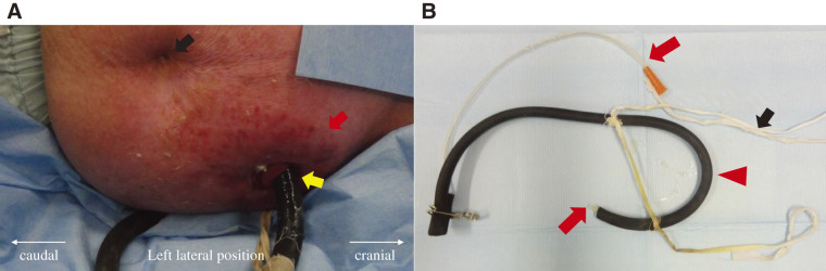
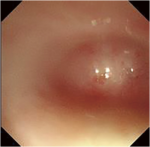
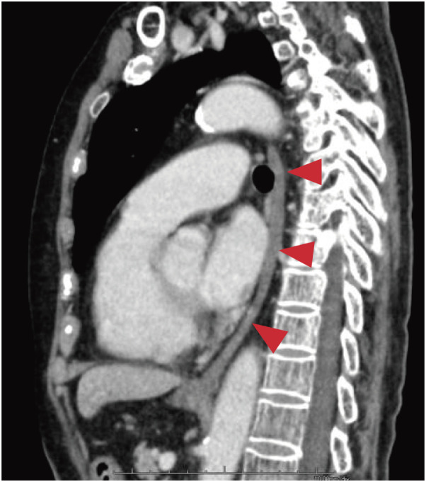
Case presentation: The patient was a 72-year-old woman. At 20 years of age, she ingested an alkali substance in a suicide attempt, leading to the development of corrosive esophagitis. Surgery was initially considered for esophageal atresia but was deemed unfeasible at the time; therefore, gastrostomy was performed instead. Subsequently, for over 50 years, she manually chewed food and inserted it into her gastric tube. She was urgently transported to a nearby hospital after her general condition deteriorated due to an influenza infection. During hospitalization, her nutritional intake was reassessed, and given her strong desire for oral intake, she was referred to our hospital for surgical treatment. Her gastric mucosa was intact, and imaging revealed mild mediastinal inflammation and fibrosis, rendering esophageal resection and reconstruction feasible. Considering surgical invasiveness, we opted for a mediastinoscopic esophagectomy and performed posterior mediastinal reconstruction using a gastric tube with a cervical hand-sewn anastomosis. The patient recovered without any complications and was discharged. Although postoperative aspiration and swallowing disorders were anticipated, the patient experienced none, likely because her unique self-feeding method preserved the functions of her masticatory and swallowing muscles.
Conclusions: We report an extremely rare case of a patient with a unique history of esophageal atresia following corrosive esophagitis for over 50 years who successfully underwent minimally invasive esophagectomy using mediastinoscopy and had a favorable outcome. Mediastinoscopic esophagectomy is a minimally invasive option for such patients.

 求助内容:
求助内容: 应助结果提醒方式:


