Sebastian Schilde, Dariusch Arbab, Maria Felsberg, Heike Kielstein, Karl-Stefan Delank, Natalia Gutteck
{"title":"Minimally Invasive Cartilage Resection of the Subtalar Joint: An Anatomical Study.","authors":"Sebastian Schilde, Dariusch Arbab, Maria Felsberg, Heike Kielstein, Karl-Stefan Delank, Natalia Gutteck","doi":"10.1177/24730114251315666","DOIUrl":null,"url":null,"abstract":"<p><strong>Background: </strong>Subtalar arthrodesis is a commonly performed procedure for the treatment of posttraumatic or primary osteoarthritis and several hindfoot deformities. The primary objective of this study is to evaluate the efficacy and safety of a minimally invasive technique for cartilage removal of the subtalar joint using a modified sinus tarsi approach.</p><p><strong>Methods: </strong>An anatomical study was performed on 8 pairs of fresh frozen cadaveric feet. A modified 2.5-cm sinus tarsi approach was used to access the subtalar joint. Cartilage removal was performed in 2 groups using either a 13-mm Shannon burr (GB) or a curette (GC) with subsequent systematic dissection. Standardized scaled photographs of the resected articular surfaces were analyzed in ImageJ software to quantify cartilage removal. Nearby vulnerable anatomical structures such as the intermediate and lateral dorsal cutaneous nerves, peroneal, tibialis posterior, flexor digitorum longus, and flexor hallucis longus tendons were assessed for injury.</p><p><strong>Results: </strong>The area of completely removed cartilage in GC was median 79.7% talar and 76.6% calcaneal. In GB, median 67.8% of the talar cartilage and 76.8% of the calcaneal cartilage was removed. The overall mean of cartilage resection was 73% (±7.7). There was no statistically significant difference between the groups. Anatomical structures at risk were not inadvertently injured.</p><p><strong>Conclusion: </strong>Subtalar cartilage resection can be performed safely using a minimally invasive modified sinus tarsi approach and either a Shannon burr or curettes. The amount of cartilage resection is less than that reported in the literature for open cartilage resection, but may be beneficial in select patient populations at increased risk for wound healing compromise and infection.</p><p><strong>Level of evidence: </strong>Level III, comparative cadaver study.</p>","PeriodicalId":12429,"journal":{"name":"Foot & Ankle Orthopaedics","volume":"10 1","pages":"24730114251315666"},"PeriodicalIF":0.0000,"publicationDate":"2025-03-19","publicationTypes":"Journal Article","fieldsOfStudy":null,"isOpenAccess":false,"openAccessPdf":"https://www.ncbi.nlm.nih.gov/pmc/articles/PMC11924069/pdf/","citationCount":"0","resultStr":null,"platform":"Semanticscholar","paperid":null,"PeriodicalName":"Foot & Ankle Orthopaedics","FirstCategoryId":"1085","ListUrlMain":"https://doi.org/10.1177/24730114251315666","RegionNum":0,"RegionCategory":null,"ArticlePicture":[],"TitleCN":null,"AbstractTextCN":null,"PMCID":null,"EPubDate":"2025/1/1 0:00:00","PubModel":"eCollection","JCR":"","JCRName":"","Score":null,"Total":0}
引用次数: 0
Abstract
Background: Subtalar arthrodesis is a commonly performed procedure for the treatment of posttraumatic or primary osteoarthritis and several hindfoot deformities. The primary objective of this study is to evaluate the efficacy and safety of a minimally invasive technique for cartilage removal of the subtalar joint using a modified sinus tarsi approach.
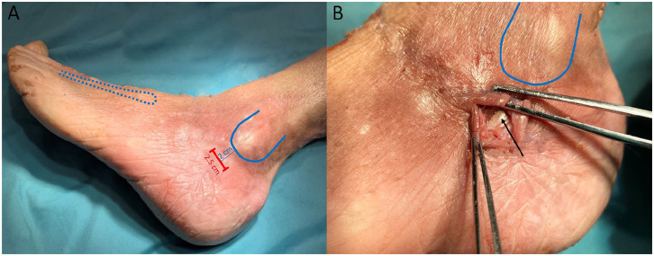
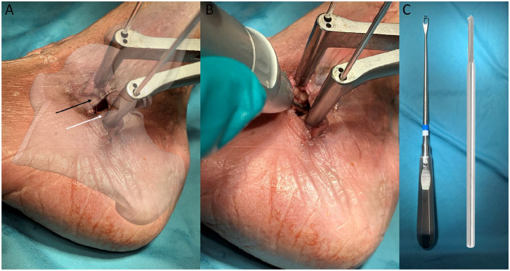
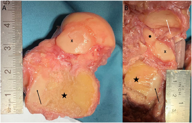
Methods: An anatomical study was performed on 8 pairs of fresh frozen cadaveric feet. A modified 2.5-cm sinus tarsi approach was used to access the subtalar joint. Cartilage removal was performed in 2 groups using either a 13-mm Shannon burr (GB) or a curette (GC) with subsequent systematic dissection. Standardized scaled photographs of the resected articular surfaces were analyzed in ImageJ software to quantify cartilage removal. Nearby vulnerable anatomical structures such as the intermediate and lateral dorsal cutaneous nerves, peroneal, tibialis posterior, flexor digitorum longus, and flexor hallucis longus tendons were assessed for injury.
Results: The area of completely removed cartilage in GC was median 79.7% talar and 76.6% calcaneal. In GB, median 67.8% of the talar cartilage and 76.8% of the calcaneal cartilage was removed. The overall mean of cartilage resection was 73% (±7.7). There was no statistically significant difference between the groups. Anatomical structures at risk were not inadvertently injured.
Conclusion: Subtalar cartilage resection can be performed safely using a minimally invasive modified sinus tarsi approach and either a Shannon burr or curettes. The amount of cartilage resection is less than that reported in the literature for open cartilage resection, but may be beneficial in select patient populations at increased risk for wound healing compromise and infection.
Level of evidence: Level III, comparative cadaver study.

 求助内容:
求助内容: 应助结果提醒方式:


