Amir Pourmorteza, Arnaud Richard Choux, Thomas Wesley Holmes, U Joseph Schoepf, Marly van Assen, Carlo De Cecco, Tilman Emrich, Akos Varga-Szemes
{"title":"Beam hardening of K-edge contrast agents: a phantom study comparing clinical energy-integrating detector and photon-counting detector CT systems.","authors":"Amir Pourmorteza, Arnaud Richard Choux, Thomas Wesley Holmes, U Joseph Schoepf, Marly van Assen, Carlo De Cecco, Tilman Emrich, Akos Varga-Szemes","doi":"10.1186/s41747-024-00530-5","DOIUrl":null,"url":null,"abstract":"<p><strong>Background: </strong>Beam hardening (BH) artifacts negatively influence computed tomography (CT) measurements, especially when due to dense materials or materials with high effective atomic numbers. Photon-counting detectors (PCD) are more susceptible to BH due to equal weighting of photons regardless of their energies. The problem is further confounded by the use of contrast agents (CAs) with K-edge in the diagnostic CT energy range. We quantified the BH effect of different materials comparing energy-integrating detector (EID)-CT and PCD-CT.</p><p><strong>Methods: </strong>Pairs of test tubes were filled with dense CA (iodine-, gadolinium-, and bismuth-based) and placed inside a water phantom. The phantoms were scanned on EID- and PCD-CT systems, at all available tube voltages for the PCD scanner. Images were reconstructed with standard water BH correction but without any iodine/bone BH corrections. Virtual monoenergetic images (VMI) were calculated from PCD-CT data.</p><p><strong>Results: </strong>PCD-CT had higher CT numbers in all x-ray spectra for all CAs (p < 0.001) and produced larger cupping artifacts in all test cases (p < 0.001). Bismuth-based CA artifacts were 3- to 5-fold smaller than those of iodine- or gadolinium-based CA. PCD-CT-based VMI completely removed iodine BH artifacts. Iodine BH artifacts decreased with increasing tube voltage. However, gadolinium-based BH artifacts had a different trend increasing at 120 kVp.</p><p><strong>Conclusion: </strong>EID had fewer BH artifacts compared to PCD at x-ray tube voltages of 120 kVp and higher. The inherent spectral information of PCDs can be used to eliminate BH artifacts. Special care is needed to correct BH artifacts for gadolinium- and bismuth-based CAs.</p><p><strong>Relevance statement: </strong>With the increasing availability of clinical photon-counting CT systems offering the possibility of dual contrast imaging capabilities, addressing and comprehending the BH artifacts attributed to old and novel CT CAs grows in research and ultimately clinical relevance.</p><p><strong>Key points: </strong>EID-CT provides fewer BH artifacts compared to PCD-CT at x-ray tube voltages of 120 kVp and higher. K-edge CAs, such as those based on gadolinium, further confound BH artifacts. The inherent spectral information of photon counting detector CT can be used to effectively eliminate BH artifacts.</p>","PeriodicalId":36926,"journal":{"name":"European Radiology Experimental","volume":"9 1","pages":"31"},"PeriodicalIF":3.6000,"publicationDate":"2025-03-19","publicationTypes":"Journal Article","fieldsOfStudy":null,"isOpenAccess":false,"openAccessPdf":"https://www.ncbi.nlm.nih.gov/pmc/articles/PMC11923337/pdf/","citationCount":"0","resultStr":null,"platform":"Semanticscholar","paperid":null,"PeriodicalName":"European Radiology Experimental","FirstCategoryId":"1085","ListUrlMain":"https://doi.org/10.1186/s41747-024-00530-5","RegionNum":0,"RegionCategory":null,"ArticlePicture":[],"TitleCN":null,"AbstractTextCN":null,"PMCID":null,"EPubDate":"","PubModel":"","JCR":"Q1","JCRName":"RADIOLOGY, NUCLEAR MEDICINE & MEDICAL IMAGING","Score":null,"Total":0}
引用次数: 0
Abstract
Background: Beam hardening (BH) artifacts negatively influence computed tomography (CT) measurements, especially when due to dense materials or materials with high effective atomic numbers. Photon-counting detectors (PCD) are more susceptible to BH due to equal weighting of photons regardless of their energies. The problem is further confounded by the use of contrast agents (CAs) with K-edge in the diagnostic CT energy range. We quantified the BH effect of different materials comparing energy-integrating detector (EID)-CT and PCD-CT.
Methods: Pairs of test tubes were filled with dense CA (iodine-, gadolinium-, and bismuth-based) and placed inside a water phantom. The phantoms were scanned on EID- and PCD-CT systems, at all available tube voltages for the PCD scanner. Images were reconstructed with standard water BH correction but without any iodine/bone BH corrections. Virtual monoenergetic images (VMI) were calculated from PCD-CT data.
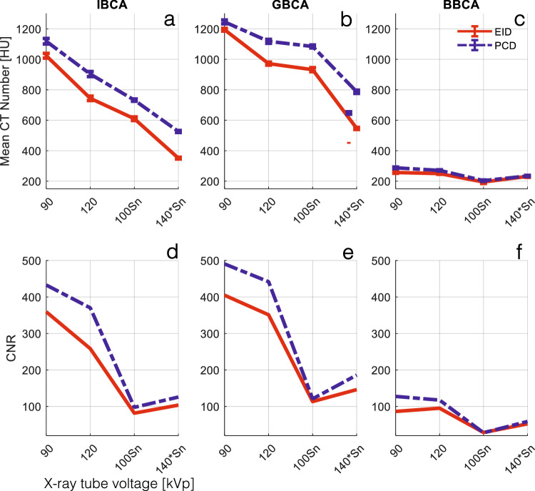
Results: PCD-CT had higher CT numbers in all x-ray spectra for all CAs (p < 0.001) and produced larger cupping artifacts in all test cases (p < 0.001). Bismuth-based CA artifacts were 3- to 5-fold smaller than those of iodine- or gadolinium-based CA. PCD-CT-based VMI completely removed iodine BH artifacts. Iodine BH artifacts decreased with increasing tube voltage. However, gadolinium-based BH artifacts had a different trend increasing at 120 kVp.
Conclusion: EID had fewer BH artifacts compared to PCD at x-ray tube voltages of 120 kVp and higher. The inherent spectral information of PCDs can be used to eliminate BH artifacts. Special care is needed to correct BH artifacts for gadolinium- and bismuth-based CAs.
Relevance statement: With the increasing availability of clinical photon-counting CT systems offering the possibility of dual contrast imaging capabilities, addressing and comprehending the BH artifacts attributed to old and novel CT CAs grows in research and ultimately clinical relevance.
Key points: EID-CT provides fewer BH artifacts compared to PCD-CT at x-ray tube voltages of 120 kVp and higher. K-edge CAs, such as those based on gadolinium, further confound BH artifacts. The inherent spectral information of photon counting detector CT can be used to effectively eliminate BH artifacts.

 求助内容:
求助内容: 应助结果提醒方式:


