{"title":"Role of Tau Protein Hyperphosphorylation in Diabetic Retinal Neurodegeneration.","authors":"Jingyu Mu, Zengrui Zhang, Chao Jiang, Haoming Geng, Junguo Duan","doi":"10.1155/joph/3278794","DOIUrl":null,"url":null,"abstract":"<p><p>Diabetic retinal neurodegeneration (DRN) is an early manifestation of diabetic retinopathy (DR) characterized by neurodegeneration that precedes microvascular abnormalities in the retina. DRN is characterized by apoptosis of retinal ganglion cells (involves alterations in retinal ganglion cells [RGCs], photoreceptors, amacrine cells and bipolar cells and so on), reactive gliosis, and reduced retinal neuronal function. Tau, a microtubule-associated protein, is a key mediator of neurotoxicity in neurodegenerative diseases, with functions in phosphorylation-dependent microtubule assembly and stabilization, axonal transport, and neurite outgrowth. The hyperphosphorylated tau (p-tau) loses its ability to bind to microtubules and aggregates to form paired helical filaments (PHFs), which further form neurofibrillary tangles (NFTs), leading to abnormal cell scaffolding and cell death. Studies have shown that p-tau can cause degeneration of RGCs in DR, making tau pathology a new pathophysiological model for DR. Here, we review the mechanisms by which p-tau contribute to DRN, including insulin resistance or lack of insulin, mitochondrial damage such as mitophagy impairment, mitochondrial axonal transport defects, mitochondrial bioenergetics dysfunction, and impaired mitochondrial dynamics, Abeta toxicity, and inflammation. Therefore, this article proposes that tau protein hyperphosphorylation plays a crucial role in the pathogenesis of DRN and may serve as a novel therapeutic target for combating DRN.</p>","PeriodicalId":16674,"journal":{"name":"Journal of Ophthalmology","volume":"2025 ","pages":"3278794"},"PeriodicalIF":1.9000,"publicationDate":"2025-03-12","publicationTypes":"Journal Article","fieldsOfStudy":null,"isOpenAccess":false,"openAccessPdf":"https://www.ncbi.nlm.nih.gov/pmc/articles/PMC11922625/pdf/","citationCount":"0","resultStr":null,"platform":"Semanticscholar","paperid":null,"PeriodicalName":"Journal of Ophthalmology","FirstCategoryId":"3","ListUrlMain":"https://doi.org/10.1155/joph/3278794","RegionNum":4,"RegionCategory":"医学","ArticlePicture":[],"TitleCN":null,"AbstractTextCN":null,"PMCID":null,"EPubDate":"2025/1/1 0:00:00","PubModel":"eCollection","JCR":"Q3","JCRName":"OPHTHALMOLOGY","Score":null,"Total":0}
引用次数: 0
Abstract
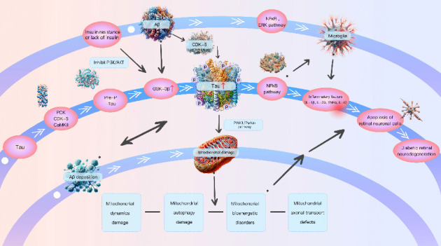
Diabetic retinal neurodegeneration (DRN) is an early manifestation of diabetic retinopathy (DR) characterized by neurodegeneration that precedes microvascular abnormalities in the retina. DRN is characterized by apoptosis of retinal ganglion cells (involves alterations in retinal ganglion cells [RGCs], photoreceptors, amacrine cells and bipolar cells and so on), reactive gliosis, and reduced retinal neuronal function. Tau, a microtubule-associated protein, is a key mediator of neurotoxicity in neurodegenerative diseases, with functions in phosphorylation-dependent microtubule assembly and stabilization, axonal transport, and neurite outgrowth. The hyperphosphorylated tau (p-tau) loses its ability to bind to microtubules and aggregates to form paired helical filaments (PHFs), which further form neurofibrillary tangles (NFTs), leading to abnormal cell scaffolding and cell death. Studies have shown that p-tau can cause degeneration of RGCs in DR, making tau pathology a new pathophysiological model for DR. Here, we review the mechanisms by which p-tau contribute to DRN, including insulin resistance or lack of insulin, mitochondrial damage such as mitophagy impairment, mitochondrial axonal transport defects, mitochondrial bioenergetics dysfunction, and impaired mitochondrial dynamics, Abeta toxicity, and inflammation. Therefore, this article proposes that tau protein hyperphosphorylation plays a crucial role in the pathogenesis of DRN and may serve as a novel therapeutic target for combating DRN.
期刊介绍:
Journal of Ophthalmology is a peer-reviewed, Open Access journal that publishes original research articles, review articles, and clinical studies related to the anatomy, physiology and diseases of the eye. Submissions should focus on new diagnostic and surgical techniques, instrument and therapy updates, as well as clinical trials and research findings.
 求助内容:
求助内容: 应助结果提醒方式:


