Daniel de Pinho Alves, Maria Eduarda Dos Santos Lopes Fernandes, Cecília Azevedo Dias Lopes, Thainá de Lima Risso, Mayara do Nascimento Trindade, Thais Marques Moreira, Isabela Scalioni Gijsen, Rodrigo Pereira da Costa Duarte
{"title":"Intraluminal unilateral ectopic ureter associated to ectopic ureterocele in a female dog - clinical, diagnostic and surgical aspects.","authors":"Daniel de Pinho Alves, Maria Eduarda Dos Santos Lopes Fernandes, Cecília Azevedo Dias Lopes, Thainá de Lima Risso, Mayara do Nascimento Trindade, Thais Marques Moreira, Isabela Scalioni Gijsen, Rodrigo Pereira da Costa Duarte","doi":"10.29374/2527-2179.bjvm008424","DOIUrl":null,"url":null,"abstract":"<p><p>Ureteral ectopia is a congenital malformation characterized by the abnormal location of the distal aspect of one or both ureters, being classified according to its anatomical path as intramural or extramural. The most common clinical sign is urinary incontinence. The presence of other associated urogenital anomalies, such as hydroureter, hydronephrosis and ureterocele are possible, being the last one a rare condition characterized as a cystic dilation of the submucosal layer of the distal ureter. The diagnosis is based on patient history, clinical signs and imaging exams. Treatment consists in surgical correction, with the technique variating according to the condition classification, and the prognosis is favorable, however most animals remain incontinent. This paper objective to report the diagnostic and clinical surgical conduction of intramural unilateral ectopic ureter correction associated to ectopic ureterocele in a Siberian Husky, 7-months-old, attended at Veterinary Hospital of Federal Rural University of Rio de Janeiro with complaint of urinary incontinence and clinical history of bacterial cystitis. The diagnosis of intramural unilateral ectopia associated with ureterocele was obtained through abdominal ultrasound and excretory urography, being confirmed with surgery. The surgical technique performed was neoureterostomy, and there were no trans or post-surgical intercurrence. Despite maintenance of the ureterocele and right ureter and renal pelvis dilation two days after surgery, observed during abdominal ultrasound, these alterations has positive evolution one week after the surgical procedure. Patient presented significant improvement of urinary incontinence two months after surgery.</p>","PeriodicalId":72458,"journal":{"name":"Brazilian journal of veterinary medicine","volume":"47 ","pages":"e008424"},"PeriodicalIF":0.0000,"publicationDate":"2025-03-18","publicationTypes":"Journal Article","fieldsOfStudy":null,"isOpenAccess":false,"openAccessPdf":"https://www.ncbi.nlm.nih.gov/pmc/articles/PMC11919245/pdf/","citationCount":"0","resultStr":null,"platform":"Semanticscholar","paperid":null,"PeriodicalName":"Brazilian journal of veterinary medicine","FirstCategoryId":"1085","ListUrlMain":"https://doi.org/10.29374/2527-2179.bjvm008424","RegionNum":0,"RegionCategory":null,"ArticlePicture":[],"TitleCN":null,"AbstractTextCN":null,"PMCID":null,"EPubDate":"2025/1/1 0:00:00","PubModel":"eCollection","JCR":"","JCRName":"","Score":null,"Total":0}
引用次数: 0
Abstract
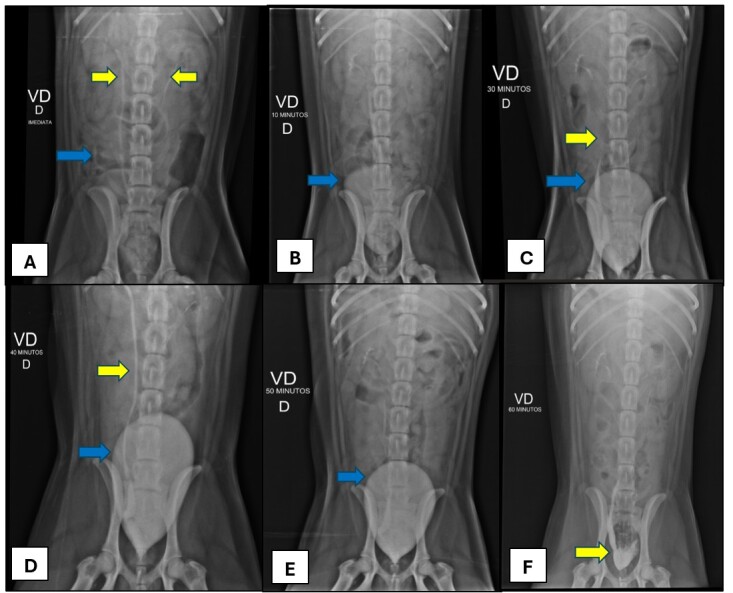
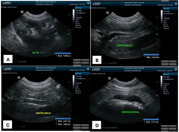
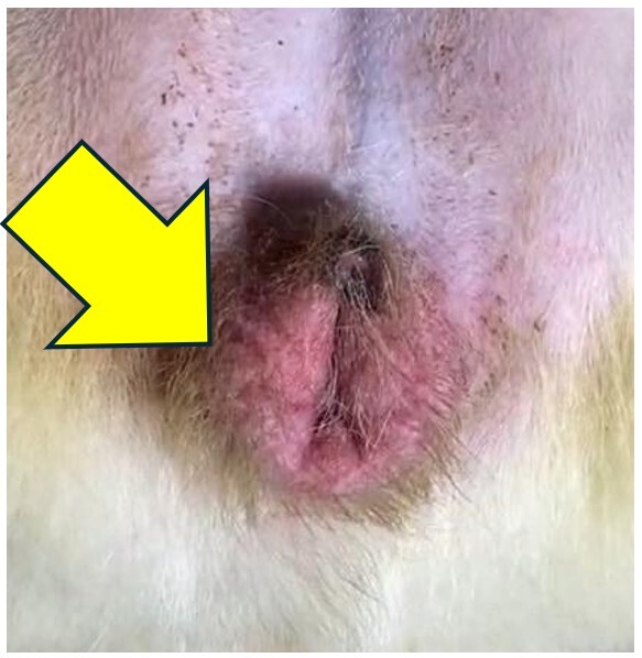
Ureteral ectopia is a congenital malformation characterized by the abnormal location of the distal aspect of one or both ureters, being classified according to its anatomical path as intramural or extramural. The most common clinical sign is urinary incontinence. The presence of other associated urogenital anomalies, such as hydroureter, hydronephrosis and ureterocele are possible, being the last one a rare condition characterized as a cystic dilation of the submucosal layer of the distal ureter. The diagnosis is based on patient history, clinical signs and imaging exams. Treatment consists in surgical correction, with the technique variating according to the condition classification, and the prognosis is favorable, however most animals remain incontinent. This paper objective to report the diagnostic and clinical surgical conduction of intramural unilateral ectopic ureter correction associated to ectopic ureterocele in a Siberian Husky, 7-months-old, attended at Veterinary Hospital of Federal Rural University of Rio de Janeiro with complaint of urinary incontinence and clinical history of bacterial cystitis. The diagnosis of intramural unilateral ectopia associated with ureterocele was obtained through abdominal ultrasound and excretory urography, being confirmed with surgery. The surgical technique performed was neoureterostomy, and there were no trans or post-surgical intercurrence. Despite maintenance of the ureterocele and right ureter and renal pelvis dilation two days after surgery, observed during abdominal ultrasound, these alterations has positive evolution one week after the surgical procedure. Patient presented significant improvement of urinary incontinence two months after surgery.

 求助内容:
求助内容: 应助结果提醒方式:


