Angela Claudia Paixão Soares de Magalhães, Gutenberg do Amaral Gurgel, Svetlana Maria Wanderley de Barros, Miguel Lucas Silva Valente, Maurício de Amorim Aquino, Sthefanie da Silva Bessa, Rogério Ferraz Baquette, Aldemar Araújo Castro, Guilherme Benjamim Brandão Pitta
{"title":"Gastrointestinal histological injury in pigs subjected to triple stent interposition in the thoracoabdominal aorta.","authors":"Angela Claudia Paixão Soares de Magalhães, Gutenberg do Amaral Gurgel, Svetlana Maria Wanderley de Barros, Miguel Lucas Silva Valente, Maurício de Amorim Aquino, Sthefanie da Silva Bessa, Rogério Ferraz Baquette, Aldemar Araújo Castro, Guilherme Benjamim Brandão Pitta","doi":"10.1590/acb402425","DOIUrl":null,"url":null,"abstract":"<p><strong>Purpose: </strong>To evaluate gastrointestinal histological injury in pigs subjected to triple stent interposition versus a control group, hypothesizing no significant injury increase with triple stents.</p><p><strong>Methods: </strong>A prospective study with 15 pigs divided into a control group (G0, n = 5) undergoing arteriography only, and a triple stent group (G3, n = 10) undergoing arteriography and three stent implantations in the thoracoabdominal aorta. After an eight-day observation, arteriography, euthanasia, and en bloc gastrointestinal harvesting were performed. Lesions were graded using the Park/Chiu classification, and serum markers were analyzed pre- and post-procedure.</p><p><strong>Results: </strong>Arteriography confirmed mesenteric artery patency in all animals. Histological analysis showed ischemic lesions in 88.9% of G3, mainly in the colon (89%), compared to 60% in G0, primarily in the colon (60%) and stomach (40%). Most G3 lesions were grade 1, while G0 had higher-grade lesions. Serum markers showed no significant intergroup differences.</p><p><strong>Conclusion: </strong>Triple stent interposition did not significantly increase gastrointestinal injury, indicating its safety for maintaining gastrointestinal perfusion in this model.</p>","PeriodicalId":93850,"journal":{"name":"Acta cirurgica brasileira","volume":"40 ","pages":"e402425"},"PeriodicalIF":1.3000,"publicationDate":"2025-03-14","publicationTypes":"Journal Article","fieldsOfStudy":null,"isOpenAccess":false,"openAccessPdf":"https://www.ncbi.nlm.nih.gov/pmc/articles/PMC11908737/pdf/","citationCount":"0","resultStr":null,"platform":"Semanticscholar","paperid":null,"PeriodicalName":"Acta cirurgica brasileira","FirstCategoryId":"1085","ListUrlMain":"https://doi.org/10.1590/acb402425","RegionNum":0,"RegionCategory":null,"ArticlePicture":[],"TitleCN":null,"AbstractTextCN":null,"PMCID":null,"EPubDate":"2025/1/1 0:00:00","PubModel":"eCollection","JCR":"","JCRName":"","Score":null,"Total":0}
引用次数: 0
Abstract
Purpose: To evaluate gastrointestinal histological injury in pigs subjected to triple stent interposition versus a control group, hypothesizing no significant injury increase with triple stents.
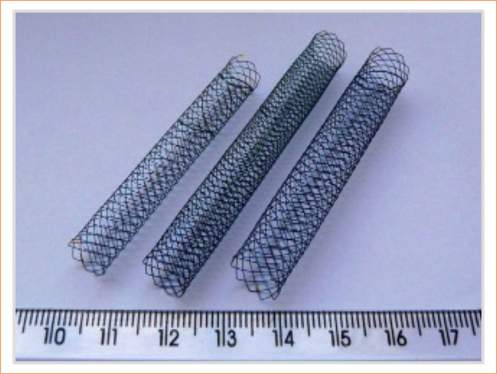
Methods: A prospective study with 15 pigs divided into a control group (G0, n = 5) undergoing arteriography only, and a triple stent group (G3, n = 10) undergoing arteriography and three stent implantations in the thoracoabdominal aorta. After an eight-day observation, arteriography, euthanasia, and en bloc gastrointestinal harvesting were performed. Lesions were graded using the Park/Chiu classification, and serum markers were analyzed pre- and post-procedure.
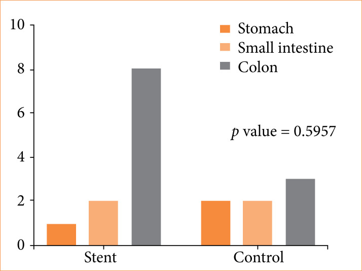
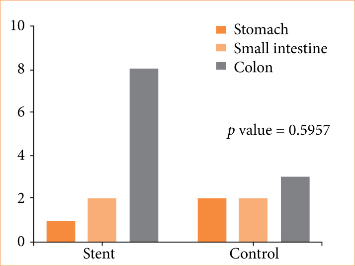
Results: Arteriography confirmed mesenteric artery patency in all animals. Histological analysis showed ischemic lesions in 88.9% of G3, mainly in the colon (89%), compared to 60% in G0, primarily in the colon (60%) and stomach (40%). Most G3 lesions were grade 1, while G0 had higher-grade lesions. Serum markers showed no significant intergroup differences.
Conclusion: Triple stent interposition did not significantly increase gastrointestinal injury, indicating its safety for maintaining gastrointestinal perfusion in this model.
目的:在假定三次支架置入没有显著损伤增加的情况下,评估三次支架置入猪与对照组的胃肠道组织学损伤。方法:采用前瞻性研究方法,将15头猪分为对照组(G0, n = 5)和三联支架组(G3, n = 10),分别在胸腹主动脉行动脉造影术和三联支架植入术。观察8天后,进行动脉造影、安乐死和整体胃肠道切除。采用Park/Chiu分级法对病变进行分级,并对术前和术后血清标志物进行分析。结果:动脉造影证实所有动物肠系膜动脉通畅。组织学分析显示,G3中缺血性病变发生率为88.9%,主要发生在结肠(89%),而G0中缺血性病变发生率为60%,主要发生在结肠(60%)和胃(40%)。大多数G3病变为1级,而G0病变级别更高。血清指标组间差异无统计学意义。结论:三联支架置入对胃肠损伤无明显增加,表明其对维持模型胃肠灌注是安全的。

 求助内容:
求助内容: 应助结果提醒方式:


