{"title":"Comparison of the effects of epidermal growth factor mesenchymal stem cell and silver sulfadiazine on burn stasis zone.","authors":"Ömer Kürklü, Sinan Soylu","doi":"10.47717/turkjsurg.2025.6684","DOIUrl":null,"url":null,"abstract":"<p><strong>Objective: </strong>This study investigates the effects of adipose tissue-derived mesenchymal stem cell (MSC), human recombinant epidermal growth factor (EGF) and silver sulfadiazine (SSD) on wound healing in the burn stasis zone by applying the comb burn model in rats.</p><p><strong>Material and methods: </strong>A comb burn model was used for the burns and 32 Wistar albino female rats were randomly divided into four groups (control, SSD, SSD+MSC, SSD+EGF). On the 1<sup>st</sup> day and the 21<sup>st</sup> day, the total burn area on the 1<sup>st</sup> day and the healed, healing, and non-healing burn area on the 21<sup>st</sup> day were calculated with the Image-J program. At the end of the 21<sup>st</sup> day, the pathology samples taken after euthanasia were scored semiquantitatively in terms of epithelization, inflammatory cell density, fibroblast density, collagen amount, and angiogenesis after hematoxylin-eosin staining.</p><p><strong>Results: </strong>Histopathological analysis demonstrated that epithelialization scores were highest in the MSC (3.88±0.35, p<0.001) and EGF (3.63±0.52) groups, while the control group had the lowest values (1.50±0.53). Inflammatory cell density was significantly lower in the MSC (1.50±0.53, p<0.001) and EGF (1.88±0.64) groups than in the control group (3.75±0.46). Similarly, fibroblast density was lowest in the MSC (1.38±0.52, p<0.001) and EGF (1.75±0.71) groups, while the control group had the highest values (3.63±0.52). Collagen fibril density was significantly increased in the MSC (3.88±0.35, p<0.001) and EGF (3.50±0.53) groups compared to the control (1.63±0.74). Angiogenesis was highest in the EGF group (3.75±0.46, p<0.001), followed by the MSC group (3.00±0.53), while the control group had the lowest values (1.25±0.46). These results suggest that MSC and EGF play a significant role in wound healing, with MSC demonstrating superior epithelialization and EGF exhibiting the greatest angiogenic effect. Photo-analytical measurements showed that on day 1, burn area sizes were similar among all groups (p>0.05). By day 21, the healing burn area was significantly smaller in the MSC (3.19±0.98 cm², p<0.001) and EGF (4.33±0.48 cm²) groups compared to the control (8.43±2.35 cm²). The non-healing area was smallest in the EGF group (0.67±0.49 cm²), followed by the MSC (1.06±0.49 cm², p<0.001) and SSD (1.91±0.75 cm²) groups, whereas the control group had the largest non-healing area (7.29±2.20 cm²). These findings suggest that MSC was the most effective treatment for promoting wound healing, followed by EGF and SSD.</p><p><strong>Conclusion: </strong>We determined that both histologically and photo analytically, MSC and EGF provided faster wound healing in the burn stasis zone EGF gave better results than all groups in preventing necrosis.</p>","PeriodicalId":23374,"journal":{"name":"Turkish Journal of Surgery","volume":" ","pages":"135-140"},"PeriodicalIF":0.6000,"publicationDate":"2025-05-30","publicationTypes":"Journal Article","fieldsOfStudy":null,"isOpenAccess":false,"openAccessPdf":"https://www.ncbi.nlm.nih.gov/pmc/articles/PMC12124341/pdf/","citationCount":"0","resultStr":null,"platform":"Semanticscholar","paperid":null,"PeriodicalName":"Turkish Journal of Surgery","FirstCategoryId":"1085","ListUrlMain":"https://doi.org/10.47717/turkjsurg.2025.6684","RegionNum":0,"RegionCategory":null,"ArticlePicture":[],"TitleCN":null,"AbstractTextCN":null,"PMCID":null,"EPubDate":"2025/3/19 0:00:00","PubModel":"Epub","JCR":"Q4","JCRName":"SURGERY","Score":null,"Total":0}
引用次数: 0
Abstract
Objective: This study investigates the effects of adipose tissue-derived mesenchymal stem cell (MSC), human recombinant epidermal growth factor (EGF) and silver sulfadiazine (SSD) on wound healing in the burn stasis zone by applying the comb burn model in rats.
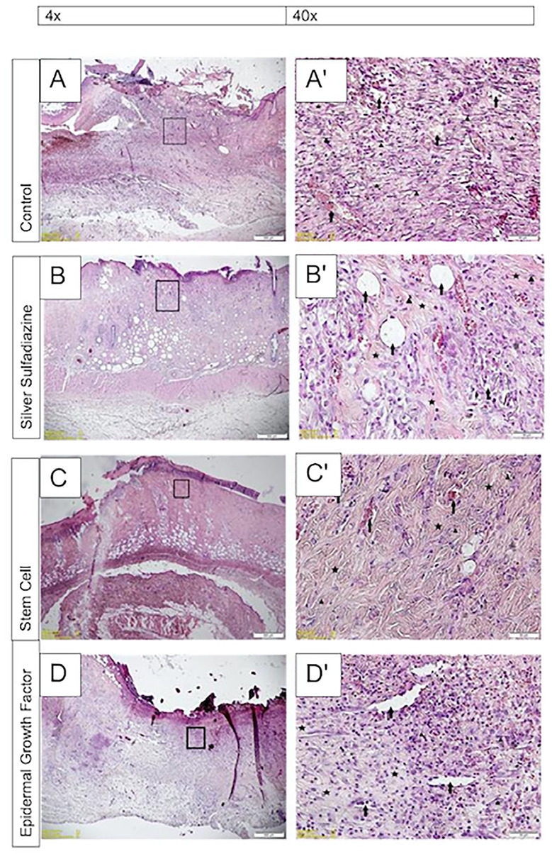
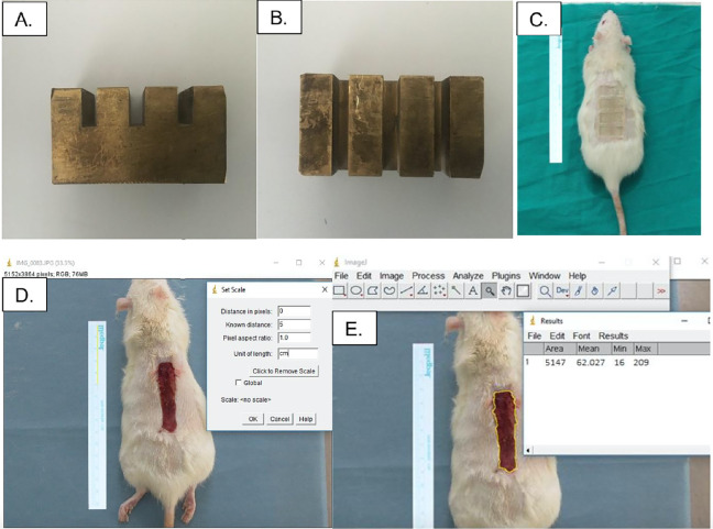
Material and methods: A comb burn model was used for the burns and 32 Wistar albino female rats were randomly divided into four groups (control, SSD, SSD+MSC, SSD+EGF). On the 1st day and the 21st day, the total burn area on the 1st day and the healed, healing, and non-healing burn area on the 21st day were calculated with the Image-J program. At the end of the 21st day, the pathology samples taken after euthanasia were scored semiquantitatively in terms of epithelization, inflammatory cell density, fibroblast density, collagen amount, and angiogenesis after hematoxylin-eosin staining.
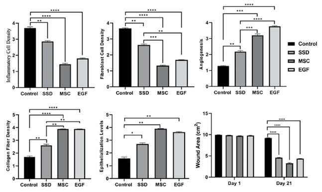
Results: Histopathological analysis demonstrated that epithelialization scores were highest in the MSC (3.88±0.35, p<0.001) and EGF (3.63±0.52) groups, while the control group had the lowest values (1.50±0.53). Inflammatory cell density was significantly lower in the MSC (1.50±0.53, p<0.001) and EGF (1.88±0.64) groups than in the control group (3.75±0.46). Similarly, fibroblast density was lowest in the MSC (1.38±0.52, p<0.001) and EGF (1.75±0.71) groups, while the control group had the highest values (3.63±0.52). Collagen fibril density was significantly increased in the MSC (3.88±0.35, p<0.001) and EGF (3.50±0.53) groups compared to the control (1.63±0.74). Angiogenesis was highest in the EGF group (3.75±0.46, p<0.001), followed by the MSC group (3.00±0.53), while the control group had the lowest values (1.25±0.46). These results suggest that MSC and EGF play a significant role in wound healing, with MSC demonstrating superior epithelialization and EGF exhibiting the greatest angiogenic effect. Photo-analytical measurements showed that on day 1, burn area sizes were similar among all groups (p>0.05). By day 21, the healing burn area was significantly smaller in the MSC (3.19±0.98 cm², p<0.001) and EGF (4.33±0.48 cm²) groups compared to the control (8.43±2.35 cm²). The non-healing area was smallest in the EGF group (0.67±0.49 cm²), followed by the MSC (1.06±0.49 cm², p<0.001) and SSD (1.91±0.75 cm²) groups, whereas the control group had the largest non-healing area (7.29±2.20 cm²). These findings suggest that MSC was the most effective treatment for promoting wound healing, followed by EGF and SSD.
Conclusion: We determined that both histologically and photo analytically, MSC and EGF provided faster wound healing in the burn stasis zone EGF gave better results than all groups in preventing necrosis.

 求助内容:
求助内容: 应助结果提醒方式:


