Renata Borges Rodrigues, Allyne Jorcelino Daloia de Carvalho, Bruna Vanessa Felipe E Silva, Paulo Cézar Simamoto-Júnior, Veridiana Resende Novais
{"title":"Impact of radiotherapy in chemical composition and mechanical properties of human cervical dentin: an in vitro study.","authors":"Renata Borges Rodrigues, Allyne Jorcelino Daloia de Carvalho, Bruna Vanessa Felipe E Silva, Paulo Cézar Simamoto-Júnior, Veridiana Resende Novais","doi":"10.1590/1678-7757-2024-0279","DOIUrl":null,"url":null,"abstract":"<p><strong>Background: </strong>Ionizing radiation directly affects hard dental tissues, compromising the dental structure, which results in damage to dentin collagen fibers and impacts the integrity of the dentin-enamel junction (DEJ).</p><p><strong>Objective: </strong>To evaluate the effects of radiotherapy on the chemical composition and mechanical properties of human cervical dentin.</p><p><strong>Methodology: </strong>Ten third molars were divided into control/non-irradiated and irradiated groups (n=5). The irradiated teeth were subjected to in vitro radiotherapy with the following protocol: 1.8 Gy daily, five days per week for eight weeks, totaling 72 Gy. The dentin in the cervical region was evaluated for each group. The chemical composition was assessed using Fourier transform infrared spectroscopy (FTIR) and Raman spectroscopy, focusing on the mineral/matrix ratio (M:M), carbonate/mineral ratio (C:M), and amide I/amide III ratio. Amide I/CH2 ratio was used to assess collagen quality, as amide I reflects protein conformation and hydrogen bonding, while CH2 indicates side-chain vibrations with low sensitivity to molecular orientation. Nanohardness and elastic modulus were evaluated by instrumented indentation. Scanning electron microscopy (SEM) was used to assess the enamel's morphology. Statistical analysis of each parameter was performed using a t-test.</p><p><strong>Results: </strong>The FTIR analysis showed statistically significant differences in the C:M ratio (p=0.004) and amide I/amide III ratio (p=0.007). Raman spectroscopy revealed significant differences in the M:M ratio (p<0.001), as well as in the amide I/amide III (p<0.001) and amide I/CH2 ratios (p<0.001). Additionally, nanohardness (p=0.04) and the elastic modulus (p=0.003) showed statistically significant differences. SEM images revealed sound dentin shows normal tissue organization, whereas irradiated dentin showed no clear limit between peri and intertubular dentin.</p><p><strong>Conclusions: </strong>Radiotherapy induced significant changes in dentin composition and mechanical properties, characterized by increased organic content and phosphate levels, reduced carbonate, and decreased nanohardness and elastic modulus. These findings highlight the adverse effects on dentin's structural integrity.</p>","PeriodicalId":15133,"journal":{"name":"Journal of Applied Oral Science","volume":"33 ","pages":"e20240279"},"PeriodicalIF":2.6000,"publicationDate":"2025-03-14","publicationTypes":"Journal Article","fieldsOfStudy":null,"isOpenAccess":false,"openAccessPdf":"https://www.ncbi.nlm.nih.gov/pmc/articles/PMC11978287/pdf/","citationCount":"0","resultStr":null,"platform":"Semanticscholar","paperid":null,"PeriodicalName":"Journal of Applied Oral Science","FirstCategoryId":"3","ListUrlMain":"https://doi.org/10.1590/1678-7757-2024-0279","RegionNum":3,"RegionCategory":"医学","ArticlePicture":[],"TitleCN":null,"AbstractTextCN":null,"PMCID":null,"EPubDate":"2025/1/1 0:00:00","PubModel":"eCollection","JCR":"Q2","JCRName":"DENTISTRY, ORAL SURGERY & MEDICINE","Score":null,"Total":0}
引用次数: 0
Abstract
Background: Ionizing radiation directly affects hard dental tissues, compromising the dental structure, which results in damage to dentin collagen fibers and impacts the integrity of the dentin-enamel junction (DEJ).
Objective: To evaluate the effects of radiotherapy on the chemical composition and mechanical properties of human cervical dentin.
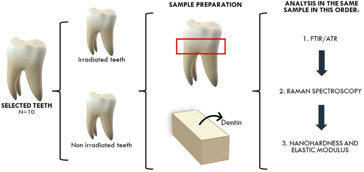
Methodology: Ten third molars were divided into control/non-irradiated and irradiated groups (n=5). The irradiated teeth were subjected to in vitro radiotherapy with the following protocol: 1.8 Gy daily, five days per week for eight weeks, totaling 72 Gy. The dentin in the cervical region was evaluated for each group. The chemical composition was assessed using Fourier transform infrared spectroscopy (FTIR) and Raman spectroscopy, focusing on the mineral/matrix ratio (M:M), carbonate/mineral ratio (C:M), and amide I/amide III ratio. Amide I/CH2 ratio was used to assess collagen quality, as amide I reflects protein conformation and hydrogen bonding, while CH2 indicates side-chain vibrations with low sensitivity to molecular orientation. Nanohardness and elastic modulus were evaluated by instrumented indentation. Scanning electron microscopy (SEM) was used to assess the enamel's morphology. Statistical analysis of each parameter was performed using a t-test.
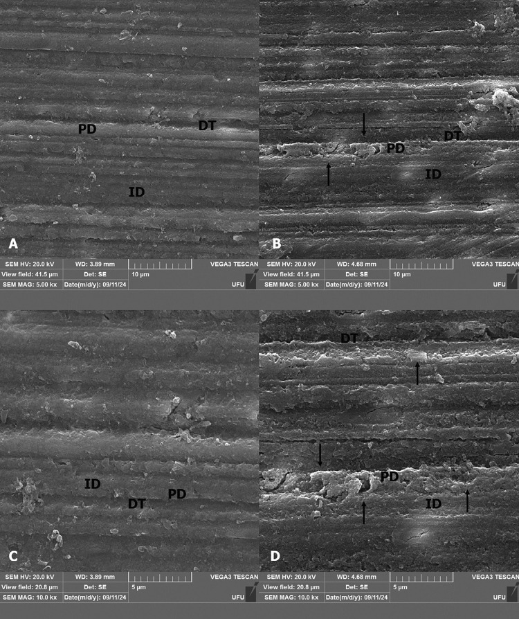
Results: The FTIR analysis showed statistically significant differences in the C:M ratio (p=0.004) and amide I/amide III ratio (p=0.007). Raman spectroscopy revealed significant differences in the M:M ratio (p<0.001), as well as in the amide I/amide III (p<0.001) and amide I/CH2 ratios (p<0.001). Additionally, nanohardness (p=0.04) and the elastic modulus (p=0.003) showed statistically significant differences. SEM images revealed sound dentin shows normal tissue organization, whereas irradiated dentin showed no clear limit between peri and intertubular dentin.
Conclusions: Radiotherapy induced significant changes in dentin composition and mechanical properties, characterized by increased organic content and phosphate levels, reduced carbonate, and decreased nanohardness and elastic modulus. These findings highlight the adverse effects on dentin's structural integrity.
期刊介绍:
The Journal of Applied Oral Science is committed in publishing the scientific and technologic advances achieved by the dental community, according to the quality indicators and peer reviewed material, with the objective of assuring its acceptability at the local, regional, national and international levels. The primary goal of The Journal of Applied Oral Science is to publish the outcomes of original investigations as well as invited case reports and invited reviews in the field of Dentistry and related areas.
 求助内容:
求助内容: 应助结果提醒方式:


