Harry Perkins, Adam B Rohrlach, Toby Hughes, Alex Forrest, Denice Higgins
{"title":"3D imaging for dental identification: a pilot investigation of a novel segmentation method using an intra oral scanning device.","authors":"Harry Perkins, Adam B Rohrlach, Toby Hughes, Alex Forrest, Denice Higgins","doi":"10.1007/s12024-025-00992-y","DOIUrl":null,"url":null,"abstract":"<p><strong>Introduction: </strong>Forensic dental identification relies on the comparison of antemortem and postmortem dental records. 3D dental imaging presents the potential for detailed anatomical features of teeth to be quantified between individuals in automated identification tools. This study introduces a novel segmentation method to simultaneously remove extraneous data from two images reducing processes and time required during 3D dental image comparisons, and tests this against existing approaches to better understand segmentation techniques for forensic purposes.</p><p><strong>Methods: </strong>Six volunteers had both digital and stone cast full arch dental models created. The casts were scanned and digitized with an intra oral laser scanner, and five different segmentation methods were then applied to all images. Segmented images were compared via a method for aligning 3D images for possible matching (same person) and non-matching (different person) pairings.</p><p><strong>Results: </strong>All segmentation methods removed adequate excess materials to provide consistent repeated outcomes in the comparison process, with the novel segmentation method showing equivalent outcomes with existing methodologies. The findings highlight the importance of understanding the process of segmentation in distinguishing between 3D dental imaging and underscore the potential of 3D imaging technologies in forensic odontology.</p><p><strong>Conclusion: </strong>The study demonstrates the efficacy of a new segmentation method in forensic dental identification, offering a faster approach; calling for further validation of these methods within a legal framework.</p>","PeriodicalId":12449,"journal":{"name":"Forensic Science, Medicine and Pathology","volume":" ","pages":"1213-1221"},"PeriodicalIF":1.4000,"publicationDate":"2025-09-01","publicationTypes":"Journal Article","fieldsOfStudy":null,"isOpenAccess":false,"openAccessPdf":"https://www.ncbi.nlm.nih.gov/pmc/articles/PMC12491115/pdf/","citationCount":"0","resultStr":null,"platform":"Semanticscholar","paperid":null,"PeriodicalName":"Forensic Science, Medicine and Pathology","FirstCategoryId":"3","ListUrlMain":"https://doi.org/10.1007/s12024-025-00992-y","RegionNum":4,"RegionCategory":"医学","ArticlePicture":[],"TitleCN":null,"AbstractTextCN":null,"PMCID":null,"EPubDate":"2025/3/18 0:00:00","PubModel":"Epub","JCR":"Q2","JCRName":"MEDICINE, LEGAL","Score":null,"Total":0}
引用次数: 0
Abstract
Introduction: Forensic dental identification relies on the comparison of antemortem and postmortem dental records. 3D dental imaging presents the potential for detailed anatomical features of teeth to be quantified between individuals in automated identification tools. This study introduces a novel segmentation method to simultaneously remove extraneous data from two images reducing processes and time required during 3D dental image comparisons, and tests this against existing approaches to better understand segmentation techniques for forensic purposes.
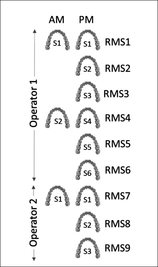
Methods: Six volunteers had both digital and stone cast full arch dental models created. The casts were scanned and digitized with an intra oral laser scanner, and five different segmentation methods were then applied to all images. Segmented images were compared via a method for aligning 3D images for possible matching (same person) and non-matching (different person) pairings.
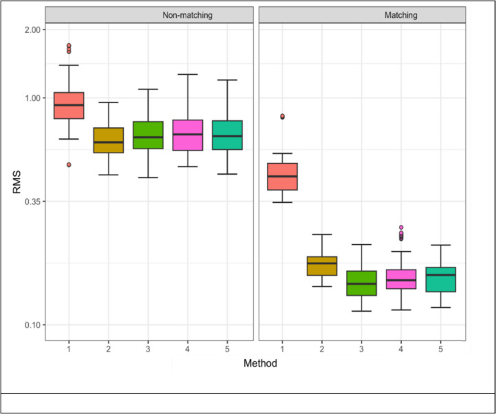
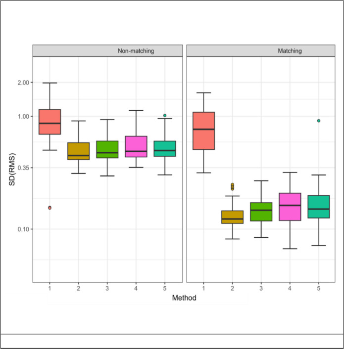
Results: All segmentation methods removed adequate excess materials to provide consistent repeated outcomes in the comparison process, with the novel segmentation method showing equivalent outcomes with existing methodologies. The findings highlight the importance of understanding the process of segmentation in distinguishing between 3D dental imaging and underscore the potential of 3D imaging technologies in forensic odontology.
Conclusion: The study demonstrates the efficacy of a new segmentation method in forensic dental identification, offering a faster approach; calling for further validation of these methods within a legal framework.
期刊介绍:
Forensic Science, Medicine and Pathology encompasses all aspects of modern day forensics, equally applying to children or adults, either living or the deceased. This includes forensic science, medicine, nursing, and pathology, as well as toxicology, human identification, mass disasters/mass war graves, profiling, imaging, policing, wound assessment, sexual assault, anthropology, archeology, forensic search, entomology, botany, biology, veterinary pathology, and DNA. Forensic Science, Medicine, and Pathology presents a balance of forensic research and reviews from around the world to reflect modern advances through peer-reviewed papers, short communications, meeting proceedings and case reports.
 求助内容:
求助内容: 应助结果提醒方式:


