François Lintz, Enrico Pozzessere, Wolfram Grün, Antoine Acker, Erik Jesús Huánuco Casas, Eric Ferkel, Cesar de Cesar Netto
{"title":"A Hallux Valgus Surgical Planning Survey Using WBCT-based 3D Printing.","authors":"François Lintz, Enrico Pozzessere, Wolfram Grün, Antoine Acker, Erik Jesús Huánuco Casas, Eric Ferkel, Cesar de Cesar Netto","doi":"10.1177/24730114251325854","DOIUrl":null,"url":null,"abstract":"<p><strong>Background: </strong>Recent literature highlights the importance of treating hallux valgus (HV) as a 3-dimensional (3D) deformity. Although 3D printing may enhance visualization of the multiplanar aspects of HV, its influence on surgical planning remains unclear. This study assessed changes in surgical plans when surgeons sequentially reviewed 2D radiographs, 3D weightbearing computed tomography (WBCT), and 3D-printed models, hypothesizing that 3D printing would have the greatest impact.</p><p><strong>Methods: </strong>A single HV case (a 40-year-old woman, intermetatarsal angle [IMA] 21 degrees, HV angle [HVA] 47 degrees) was evaluated by 30 surgeons in a masked, stepwise manner. Surgical plans were recorded at each stage. Surgeons rated the influence of WBCT and 3D printing using a 5-point Likert scale. A follow-up survey examined the effect of these technologies on correction amplitudes.</p><p><strong>Results: </strong>The participants were mostly early career surgeons (median age 35.5 years, 2 years in practice). WBCT was accessible to 43.3% and used in 30% of HV cases, whereas 3D printing was accessible to 23.3% and used in 6.6%. Changes in the treatment algorithm occurred in 30% of cases after WBCT and in 43.3% after 3D printing. Significant differences (<i>P</i> < .05) were observed for the Lapicotton procedure between radiography and WBCT, and between WBCT and 3D printing. Surgeons performing <50 HV cases annually or with >70% Foot and Ankle specialization were more influenced by WBCT. Follow-up data (n = 23) indicated that WBCT and 3D printing influenced correction amplitudes, particularly for pronation and distal metatarsal articular angle (DMAA), more than for the IMA.</p><p><strong>Discussion: </strong>Both WBCT and 3D printing influenced surgical planning, mostly explained by changes in first ray tarsometatarsal procedures. The rotational components (pronation and DMAA) were perceived as the most significantly affected. Future studies should explore cost-effectiveness, patient outcomes, and the utility of combining WBCT and 3D printing in other deformities requiring multiplanar corrections.<b>Level of Evidence:</b> Level IV, cross-sectional survey.</p>","PeriodicalId":12429,"journal":{"name":"Foot & Ankle Orthopaedics","volume":"10 1","pages":"24730114251325854"},"PeriodicalIF":0.0000,"publicationDate":"2025-03-18","publicationTypes":"Journal Article","fieldsOfStudy":null,"isOpenAccess":false,"openAccessPdf":"https://www.ncbi.nlm.nih.gov/pmc/articles/PMC11915313/pdf/","citationCount":"0","resultStr":null,"platform":"Semanticscholar","paperid":null,"PeriodicalName":"Foot & Ankle Orthopaedics","FirstCategoryId":"1085","ListUrlMain":"https://doi.org/10.1177/24730114251325854","RegionNum":0,"RegionCategory":null,"ArticlePicture":[],"TitleCN":null,"AbstractTextCN":null,"PMCID":null,"EPubDate":"2025/1/1 0:00:00","PubModel":"eCollection","JCR":"","JCRName":"","Score":null,"Total":0}
引用次数: 0
Abstract
Background: Recent literature highlights the importance of treating hallux valgus (HV) as a 3-dimensional (3D) deformity. Although 3D printing may enhance visualization of the multiplanar aspects of HV, its influence on surgical planning remains unclear. This study assessed changes in surgical plans when surgeons sequentially reviewed 2D radiographs, 3D weightbearing computed tomography (WBCT), and 3D-printed models, hypothesizing that 3D printing would have the greatest impact.
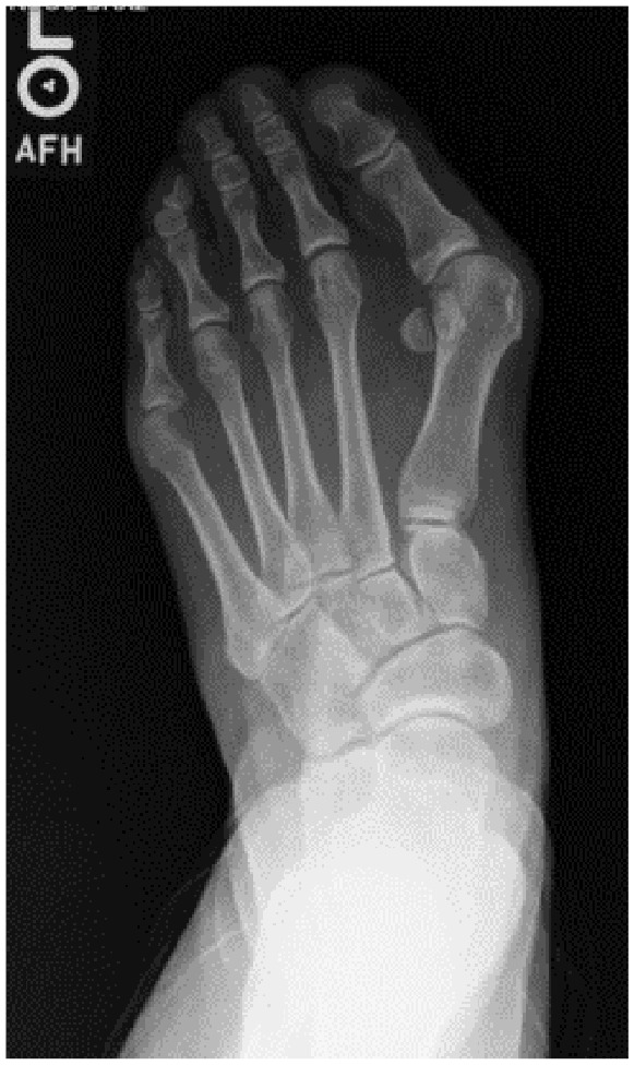
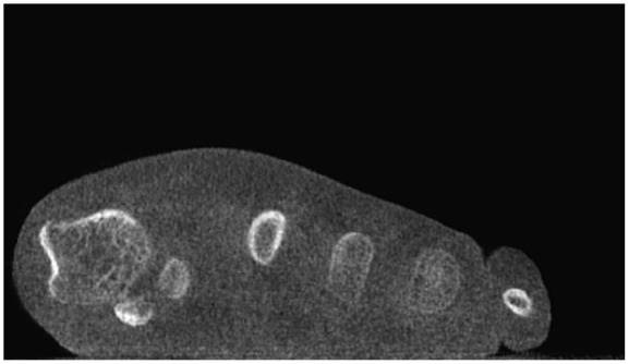
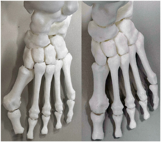
Methods: A single HV case (a 40-year-old woman, intermetatarsal angle [IMA] 21 degrees, HV angle [HVA] 47 degrees) was evaluated by 30 surgeons in a masked, stepwise manner. Surgical plans were recorded at each stage. Surgeons rated the influence of WBCT and 3D printing using a 5-point Likert scale. A follow-up survey examined the effect of these technologies on correction amplitudes.
Results: The participants were mostly early career surgeons (median age 35.5 years, 2 years in practice). WBCT was accessible to 43.3% and used in 30% of HV cases, whereas 3D printing was accessible to 23.3% and used in 6.6%. Changes in the treatment algorithm occurred in 30% of cases after WBCT and in 43.3% after 3D printing. Significant differences (P < .05) were observed for the Lapicotton procedure between radiography and WBCT, and between WBCT and 3D printing. Surgeons performing <50 HV cases annually or with >70% Foot and Ankle specialization were more influenced by WBCT. Follow-up data (n = 23) indicated that WBCT and 3D printing influenced correction amplitudes, particularly for pronation and distal metatarsal articular angle (DMAA), more than for the IMA.
Discussion: Both WBCT and 3D printing influenced surgical planning, mostly explained by changes in first ray tarsometatarsal procedures. The rotational components (pronation and DMAA) were perceived as the most significantly affected. Future studies should explore cost-effectiveness, patient outcomes, and the utility of combining WBCT and 3D printing in other deformities requiring multiplanar corrections.Level of Evidence: Level IV, cross-sectional survey.

 求助内容:
求助内容: 应助结果提醒方式:


