Dean Girotto, Josip Burić, Hrvoje Šimić, Vjekoslav Tomulić, Tomislav Jakljević, Zvonimir Kvas
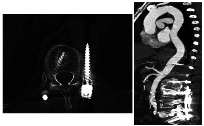
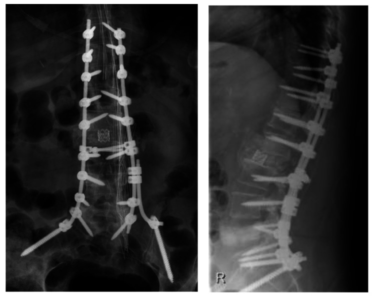
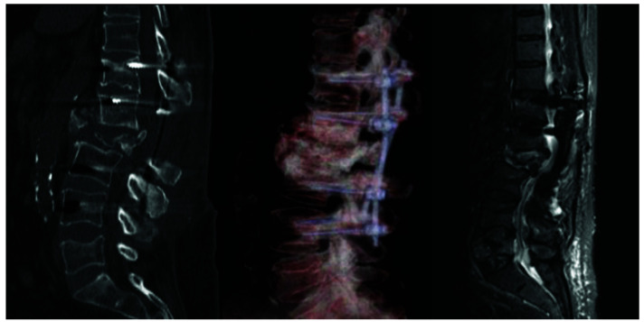
{"title":"COMBINED ENDOVASCULAR REPAIR OF AORTA AND REMOVAL OF PENETRATING PEDICLE SCREW AFTER POSTERIOR INSTRUMENTATION: A CASE REPORT AND LITERATURE REVIEW.","authors":"Dean Girotto, Josip Burić, Hrvoje Šimić, Vjekoslav Tomulić, Tomislav Jakljević, Zvonimir Kvas","doi":"10.20471/acc.2024.63.02.20","DOIUrl":null,"url":null,"abstract":"<p><p>Stabilization of spine using transpedicular screws is the most commonly used instrumentation technique among spinal surgeons. The 'free hand' technique is considered relatively safe and can be performed under x-ray control. Vascular injuries with misplaced screws are rare but potentially fatal complications. Injury of thoracoabdominal aorta by malpositioned screw demands a multidisciplinary approach. Injury of vessel wall might demand screw removal and vessel wall repair. Here we present a case of 72-year-old female patient who underwent long segment fixation of thoracolumbar spine. During follow up, computed tomography (CT) scan and afterwards aortography showed a lesion of the posterior aortic wall by malpositioned screw without signs of bleeding. After meticulous preparation, combined endovascular repair with stent-graft and removal of the penetrating screw were performed. Endovascular treatment was performed simultaneously with screw removal. During screw removal, the patient was in lateral decubital position. The patient was discharged on postoperative day 8. Follow up CT aortography 6 months later showed no leak or other changes in the aorta. We found combined endovascular vessel repair with simultaneous screw removal safe and sufficient for this kind of aortic injury. Although lateral decubital position bears limitations, it gives enough space for the operator. Performing intraoperative aortography provides good insight into stent position and possible bleeding after screw removal.</p>","PeriodicalId":7072,"journal":{"name":"Acta clinica Croatica","volume":"63 2","pages":"422-430"},"PeriodicalIF":0.8000,"publicationDate":"2024-10-01","publicationTypes":"Journal Article","fieldsOfStudy":null,"isOpenAccess":false,"openAccessPdf":"https://www.ncbi.nlm.nih.gov/pmc/articles/PMC11912848/pdf/","citationCount":"0","resultStr":null,"platform":"Semanticscholar","paperid":null,"PeriodicalName":"Acta clinica Croatica","FirstCategoryId":"3","ListUrlMain":"https://doi.org/10.20471/acc.2024.63.02.20","RegionNum":4,"RegionCategory":"医学","ArticlePicture":[],"TitleCN":null,"AbstractTextCN":null,"PMCID":null,"EPubDate":"","PubModel":"","JCR":"Q3","JCRName":"MEDICINE, GENERAL & INTERNAL","Score":null,"Total":0}
引用次数: 0
Abstract
Stabilization of spine using transpedicular screws is the most commonly used instrumentation technique among spinal surgeons. The 'free hand' technique is considered relatively safe and can be performed under x-ray control. Vascular injuries with misplaced screws are rare but potentially fatal complications. Injury of thoracoabdominal aorta by malpositioned screw demands a multidisciplinary approach. Injury of vessel wall might demand screw removal and vessel wall repair. Here we present a case of 72-year-old female patient who underwent long segment fixation of thoracolumbar spine. During follow up, computed tomography (CT) scan and afterwards aortography showed a lesion of the posterior aortic wall by malpositioned screw without signs of bleeding. After meticulous preparation, combined endovascular repair with stent-graft and removal of the penetrating screw were performed. Endovascular treatment was performed simultaneously with screw removal. During screw removal, the patient was in lateral decubital position. The patient was discharged on postoperative day 8. Follow up CT aortography 6 months later showed no leak or other changes in the aorta. We found combined endovascular vessel repair with simultaneous screw removal safe and sufficient for this kind of aortic injury. Although lateral decubital position bears limitations, it gives enough space for the operator. Performing intraoperative aortography provides good insight into stent position and possible bleeding after screw removal.
期刊介绍:
Acta Clinica Croatica is a peer reviewed general medical journal that publishes original articles that advance and improve medical science and practice and that serve the purpose of transfer of original and valuable information to journal readers. Acta Clinica Croatica is published in English four times a year.
 求助内容:
求助内容: 应助结果提醒方式:


