{"title":"Value of Ultrasonography in Assessment of Screen-Detected Calcified Ductal Carcinoma <i>In Situ</i>: A Clinical Audit.","authors":"Ukamaka Dorothy Itanyi, Deborah Allen, Ivy Okereke","doi":"10.4103/jwas.jwas_8_24","DOIUrl":null,"url":null,"abstract":"<p><strong>Background: </strong>Approximately 80% of ductal carcinoma <i>in situ</i> (DCIS) cases are asymptomatic and manifest as microcalcifications, usually detected on screening mammograms. Stereotactic biopsy is used as the primary modality for histopathologic diagnosis. Ultrasonography has been relied upon as a potentially viable alternative for early breast cancer detection, especially in low- and medium-income countries.</p><p><strong>Aim: </strong>Determine the role and reliability of breast ultrasonography in the evaluation of screen-detected microcalcifications using histologically proven calcified DCIS as a case study.</p><p><strong>Materials and methods: </strong>Cross-sectional evaluation of records of patients recalled for M3-M5 (indeterminate to malignant) type microcalcifications on screening mammograms, with a histological diagnosis of pure DCIS, in an NHS Trust Hospital, Kent, England, from March 2021 to April 2023.</p><p><strong>Results: </strong>Eighty women included were within the age range of 46-79 years, with mean age of 60 years. Background breast density was predominantly fatty in 51/80 (63.8%), without any correlation with mammographic abnormalities. Microcalcifications were classified as indeterminate (M3) in 42/80 (52.5%), while suspicious (M4) and malignant (M5) types were seen in 38/80 (47.5%). No targeted sonographic abnormalities were noted in 69 (86.2%) of the patients, while 11 (13.8%) had sonographic abnormalities. The predominant sonographic feature was non-mass hypoechogenicity in 6/11(55%). There was a correlation between mammographic code, lesion size, and ultrasonic abnormality, with 9/11 (81.8%) patients with sonographic lesions having suspicious and malignant type calcifications. The mean mammographic lesion size was significantly greater in women with abnormal ultrasound findings, 42 vs. 20 mm. Histological tumour grade was high grade in 10/11 (90.9%) lesions.</p><p><strong>Conclusion: </strong>The accuracy of breast ultrasonography as an adjunct in the detection of screen-detected microcalcifications and subsequent guided biopsy is higher when dealing with malignant type microcalcifications >15 mm in size.</p>","PeriodicalId":73993,"journal":{"name":"Journal of the West African College of Surgeons","volume":"15 2","pages":"164-170"},"PeriodicalIF":0.0000,"publicationDate":"2025-04-01","publicationTypes":"Journal Article","fieldsOfStudy":null,"isOpenAccess":false,"openAccessPdf":"https://www.ncbi.nlm.nih.gov/pmc/articles/PMC11908716/pdf/","citationCount":"0","resultStr":null,"platform":"Semanticscholar","paperid":null,"PeriodicalName":"Journal of the West African College of Surgeons","FirstCategoryId":"1085","ListUrlMain":"https://doi.org/10.4103/jwas.jwas_8_24","RegionNum":0,"RegionCategory":null,"ArticlePicture":[],"TitleCN":null,"AbstractTextCN":null,"PMCID":null,"EPubDate":"2024/9/2 0:00:00","PubModel":"Epub","JCR":"","JCRName":"","Score":null,"Total":0}
引用次数: 0
Abstract
Background: Approximately 80% of ductal carcinoma in situ (DCIS) cases are asymptomatic and manifest as microcalcifications, usually detected on screening mammograms. Stereotactic biopsy is used as the primary modality for histopathologic diagnosis. Ultrasonography has been relied upon as a potentially viable alternative for early breast cancer detection, especially in low- and medium-income countries.
Aim: Determine the role and reliability of breast ultrasonography in the evaluation of screen-detected microcalcifications using histologically proven calcified DCIS as a case study.
Materials and methods: Cross-sectional evaluation of records of patients recalled for M3-M5 (indeterminate to malignant) type microcalcifications on screening mammograms, with a histological diagnosis of pure DCIS, in an NHS Trust Hospital, Kent, England, from March 2021 to April 2023.
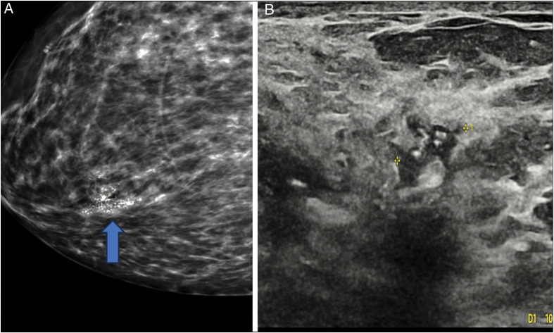
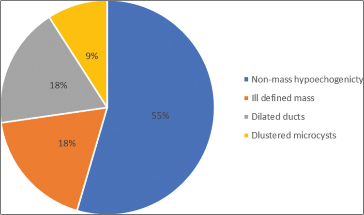
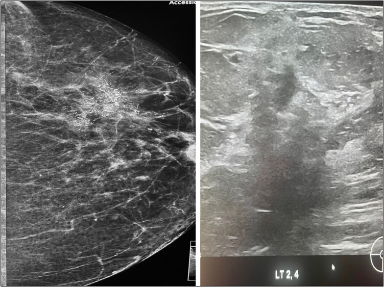
Results: Eighty women included were within the age range of 46-79 years, with mean age of 60 years. Background breast density was predominantly fatty in 51/80 (63.8%), without any correlation with mammographic abnormalities. Microcalcifications were classified as indeterminate (M3) in 42/80 (52.5%), while suspicious (M4) and malignant (M5) types were seen in 38/80 (47.5%). No targeted sonographic abnormalities were noted in 69 (86.2%) of the patients, while 11 (13.8%) had sonographic abnormalities. The predominant sonographic feature was non-mass hypoechogenicity in 6/11(55%). There was a correlation between mammographic code, lesion size, and ultrasonic abnormality, with 9/11 (81.8%) patients with sonographic lesions having suspicious and malignant type calcifications. The mean mammographic lesion size was significantly greater in women with abnormal ultrasound findings, 42 vs. 20 mm. Histological tumour grade was high grade in 10/11 (90.9%) lesions.
Conclusion: The accuracy of breast ultrasonography as an adjunct in the detection of screen-detected microcalcifications and subsequent guided biopsy is higher when dealing with malignant type microcalcifications >15 mm in size.

 求助内容:
求助内容: 应助结果提醒方式:


