Farhad Ghorbani, Mohammad Amin Amiri, Mohammad Mehdi Farahmand, Saeed Afzoun, Maryam Paknahad
{"title":"Calcifying Odontogenic Cyst in Posterior Maxilla: A Case Report.","authors":"Farhad Ghorbani, Mohammad Amin Amiri, Mohammad Mehdi Farahmand, Saeed Afzoun, Maryam Paknahad","doi":"10.30476/dentjods.2024.102636.2375","DOIUrl":null,"url":null,"abstract":"<p><p>In the present study, we reported a 66-year-old woman with an uncommonly painful calcifying odontogenic cyst (COC) in the posterior region of the left side of the maxilla. The cyst was evaluated radiographically and histopathologically. The present case showed a multilocular cyst with a mixed internal structure. The most noticeable effects on the peripheral structures were elevated maxillary sinus floor, osteomeatal complex, and nasal obstruction. To better understand the impact of COC on the trabecular pattern of the surrounding bone, we performed fractal analysis on the panoramic images pre- and post-operatively. The expansion of COC can change the trabecular pattern, which subsequently can change the fractal dimension of the area. After histopathological confirmation of the diagnosis, the cyst was surgically removed.</p>","PeriodicalId":73702,"journal":{"name":"Journal of dentistry (Shiraz, Iran)","volume":"26 1","pages":"88-94"},"PeriodicalIF":0.0000,"publicationDate":"2025-03-01","publicationTypes":"Journal Article","fieldsOfStudy":null,"isOpenAccess":false,"openAccessPdf":"https://www.ncbi.nlm.nih.gov/pmc/articles/PMC11909402/pdf/","citationCount":"0","resultStr":null,"platform":"Semanticscholar","paperid":null,"PeriodicalName":"Journal of dentistry (Shiraz, Iran)","FirstCategoryId":"1085","ListUrlMain":"https://doi.org/10.30476/dentjods.2024.102636.2375","RegionNum":0,"RegionCategory":null,"ArticlePicture":[],"TitleCN":null,"AbstractTextCN":null,"PMCID":null,"EPubDate":"","PubModel":"","JCR":"","JCRName":"","Score":null,"Total":0}
引用次数: 0
Abstract
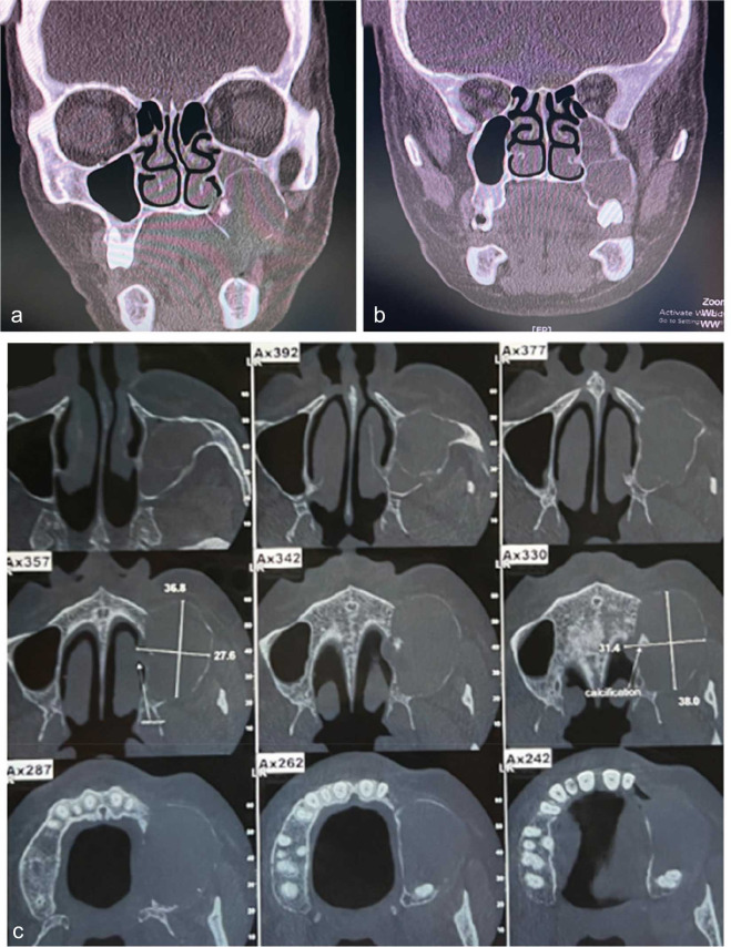
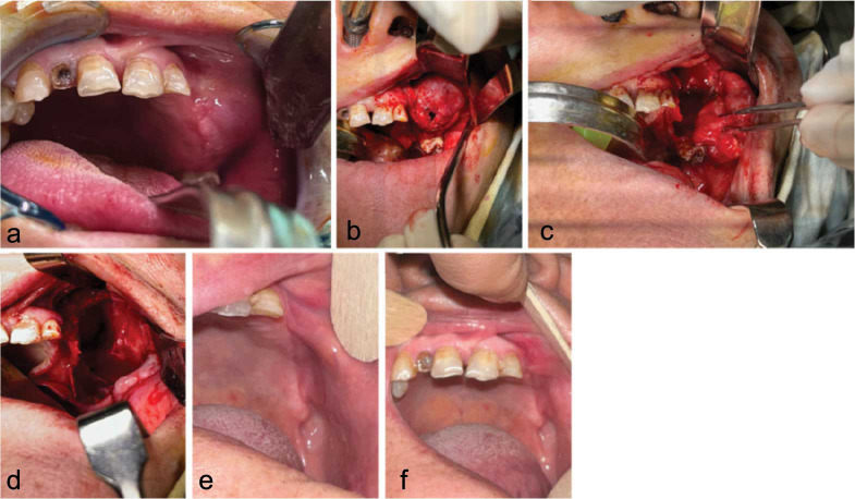
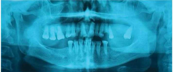
In the present study, we reported a 66-year-old woman with an uncommonly painful calcifying odontogenic cyst (COC) in the posterior region of the left side of the maxilla. The cyst was evaluated radiographically and histopathologically. The present case showed a multilocular cyst with a mixed internal structure. The most noticeable effects on the peripheral structures were elevated maxillary sinus floor, osteomeatal complex, and nasal obstruction. To better understand the impact of COC on the trabecular pattern of the surrounding bone, we performed fractal analysis on the panoramic images pre- and post-operatively. The expansion of COC can change the trabecular pattern, which subsequently can change the fractal dimension of the area. After histopathological confirmation of the diagnosis, the cyst was surgically removed.

 求助内容:
求助内容: 应助结果提醒方式:


