Joo-Young Lee, Jihye Choi, Seung-Jo Park, Jin-Woo Jung, Munsu Yun, Seonghyeon Baek, Kija Lee, Sang-Kwon Lee
{"title":"Focal Dorsal Enhancement of the Tympanic Bulla: An Early Indicator in Feline Otitis Media.","authors":"Joo-Young Lee, Jihye Choi, Seung-Jo Park, Jin-Woo Jung, Munsu Yun, Seonghyeon Baek, Kija Lee, Sang-Kwon Lee","doi":"10.1111/vru.70021","DOIUrl":null,"url":null,"abstract":"<p><p>Evaluating the morphological changes of the tympanic bulla and the contrast enhancement (CE) patterns are key factors in diagnosing otitis media and predicting its underlying etiology. However, limited research exists on the CE patterns of the tympanic bulla in cats with bulla effusion. This retrospective study aimed to investigate the prevalence and patterns of CE in the tympanic bulla based on the presence and severity of bulla effusion in cats. Feline head CT or brain MRI images from six institutions were analyzed. Transverse pre- and postcontrast CT and MRI images were reviewed to assess the presence and severity of bulla effusion, along with the presence, pattern, and location of tympanic bulla CE. A total of 644 tympanic bullae from 322 cats were included. Bulla effusion was detected in 105 of 644 bullae, while CE was observed in 73 of 644 bullae: 5 of 539 bullae without effusion and 68 of 105 bullae with effusion. CE was significantly more common in cats with bulla effusion, with the incidence increasing as effusion severity progressed. Focal rim enhancement, predominantly localized to the dorsal aspect of the tympanic bulla, was the most frequent enhancement pattern. In cases with mild or moderate bulla effusion, only focal dorsal enhancement was observed. However, in cats with severe bulla effusion, additional patterns, including focal ventral or lateral, complete rim, and internal enhancement, emerged. These findings suggest that both the incidence and patterns of CE evolve with the progression of bulla effusion in cats and that focal dorsal enhancement may be an early indicator.</p>","PeriodicalId":23581,"journal":{"name":"Veterinary Radiology & Ultrasound","volume":"66 2","pages":"e70021"},"PeriodicalIF":1.5000,"publicationDate":"2025-03-01","publicationTypes":"Journal Article","fieldsOfStudy":null,"isOpenAccess":false,"openAccessPdf":"https://www.ncbi.nlm.nih.gov/pmc/articles/PMC11911501/pdf/","citationCount":"0","resultStr":null,"platform":"Semanticscholar","paperid":null,"PeriodicalName":"Veterinary Radiology & Ultrasound","FirstCategoryId":"97","ListUrlMain":"https://doi.org/10.1111/vru.70021","RegionNum":2,"RegionCategory":"农林科学","ArticlePicture":[],"TitleCN":null,"AbstractTextCN":null,"PMCID":null,"EPubDate":"","PubModel":"","JCR":"Q2","JCRName":"VETERINARY SCIENCES","Score":null,"Total":0}
引用次数: 0
Abstract
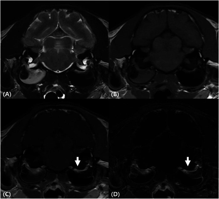
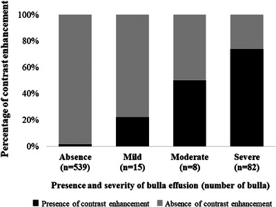
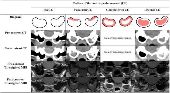
Evaluating the morphological changes of the tympanic bulla and the contrast enhancement (CE) patterns are key factors in diagnosing otitis media and predicting its underlying etiology. However, limited research exists on the CE patterns of the tympanic bulla in cats with bulla effusion. This retrospective study aimed to investigate the prevalence and patterns of CE in the tympanic bulla based on the presence and severity of bulla effusion in cats. Feline head CT or brain MRI images from six institutions were analyzed. Transverse pre- and postcontrast CT and MRI images were reviewed to assess the presence and severity of bulla effusion, along with the presence, pattern, and location of tympanic bulla CE. A total of 644 tympanic bullae from 322 cats were included. Bulla effusion was detected in 105 of 644 bullae, while CE was observed in 73 of 644 bullae: 5 of 539 bullae without effusion and 68 of 105 bullae with effusion. CE was significantly more common in cats with bulla effusion, with the incidence increasing as effusion severity progressed. Focal rim enhancement, predominantly localized to the dorsal aspect of the tympanic bulla, was the most frequent enhancement pattern. In cases with mild or moderate bulla effusion, only focal dorsal enhancement was observed. However, in cats with severe bulla effusion, additional patterns, including focal ventral or lateral, complete rim, and internal enhancement, emerged. These findings suggest that both the incidence and patterns of CE evolve with the progression of bulla effusion in cats and that focal dorsal enhancement may be an early indicator.
期刊介绍:
Veterinary Radiology & Ultrasound is a bimonthly, international, peer-reviewed, research journal devoted to the fields of veterinary diagnostic imaging and radiation oncology. Established in 1958, it is owned by the American College of Veterinary Radiology and is also the official journal for six affiliate veterinary organizations. Veterinary Radiology & Ultrasound is represented on the International Committee of Medical Journal Editors, World Association of Medical Editors, and Committee on Publication Ethics.
The mission of Veterinary Radiology & Ultrasound is to serve as a leading resource for high quality articles that advance scientific knowledge and standards of clinical practice in the areas of veterinary diagnostic radiology, computed tomography, magnetic resonance imaging, ultrasonography, nuclear imaging, radiation oncology, and interventional radiology. Manuscript types include original investigations, imaging diagnosis reports, review articles, editorials and letters to the Editor. Acceptance criteria include originality, significance, quality, reader interest, composition and adherence to author guidelines.

 求助内容:
求助内容: 应助结果提醒方式:


