Plern Sutra, Thananop Pothikamjorn, Sarah Lopez, Jaskirat Takhar, Mathinee Chongchareon, Jeremy Keenan, John A Gonzales
{"title":"Insights into scleral violaceous hue in anterior scleritis: anterior segment optical coherence tomography evaluation.","authors":"Plern Sutra, Thananop Pothikamjorn, Sarah Lopez, Jaskirat Takhar, Mathinee Chongchareon, Jeremy Keenan, John A Gonzales","doi":"10.1007/s00417-025-06788-8","DOIUrl":null,"url":null,"abstract":"<p><strong>Purpose: </strong>To determine the scleral thickness of inactive scleritis characterized by a violaceous hue (violaceous sclera) using anterior segment optical coherence tomography (AS-OCT).</p><p><strong>Methods: </strong>Retrospective observational case series of patients with inactive unilateral anterior scleritis featuring a violaceous hue. Mean scleral thickness was measured by AS-OCT in violaceous areas and compared with the same region in the contralateral unaffected eye. Measurements were performed by two masked graders.</p><p><strong>Results: </strong>Nine patients with median age of 52 ± 12.8 years were assessed. Eight patients were female. Rheumatoid arthritis and history of treated latent tuberculosis (33.3%) were the most common causes of anterior scleritis. Mean scleral thickness was 582.93 ± 124.03 µm and 648.59 ± 103.61 µm for violaceous sclera and the corresponding unaffected areas of the contralateral eye, respectively (mean difference = -65.65 µm, 95% CI: -143.73 to 12.42, p = 0.0885). The mean image contrast percentage of scleral hyperreflectivity as assessed by image conversion in an area of violaceous hue was 65.07 µm ± 6.49 µm compared to 42.70 µm ± 5.46 µm of unaffected areas (mean difference = 22.37 µm, 95% CI: 14.72 µm to 30.03 µm, p = 0.0001).</p><p><strong>Conclusion: </strong>Using AS-OCT, the thicknesses of violaceous sclerae were not significantly thinner than the contralateral unaffected areas, despite a mean difference of approximately 65 microns. The increased scleral hyperreflectivity observed in the violaceous sclera may suggest collagen remodeling in these areas. Such remodeling could play a role in the sclera reflecting violaceous hues while still preventing direct visualization of the underlying choroid.</p>","PeriodicalId":12795,"journal":{"name":"Graefe’s Archive for Clinical and Experimental Ophthalmology","volume":" ","pages":"1997-2004"},"PeriodicalIF":2.4000,"publicationDate":"2025-07-01","publicationTypes":"Journal Article","fieldsOfStudy":null,"isOpenAccess":false,"openAccessPdf":"https://www.ncbi.nlm.nih.gov/pmc/articles/PMC12373549/pdf/","citationCount":"0","resultStr":null,"platform":"Semanticscholar","paperid":null,"PeriodicalName":"Graefe’s Archive for Clinical and Experimental Ophthalmology","FirstCategoryId":"3","ListUrlMain":"https://doi.org/10.1007/s00417-025-06788-8","RegionNum":3,"RegionCategory":"医学","ArticlePicture":[],"TitleCN":null,"AbstractTextCN":null,"PMCID":null,"EPubDate":"2025/3/17 0:00:00","PubModel":"Epub","JCR":"Q2","JCRName":"OPHTHALMOLOGY","Score":null,"Total":0}
引用次数: 0
Abstract
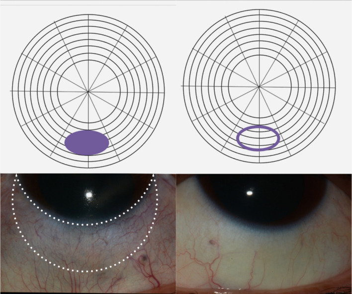
Purpose: To determine the scleral thickness of inactive scleritis characterized by a violaceous hue (violaceous sclera) using anterior segment optical coherence tomography (AS-OCT).
Methods: Retrospective observational case series of patients with inactive unilateral anterior scleritis featuring a violaceous hue. Mean scleral thickness was measured by AS-OCT in violaceous areas and compared with the same region in the contralateral unaffected eye. Measurements were performed by two masked graders.
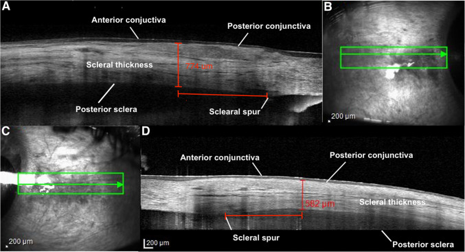
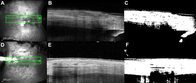
Results: Nine patients with median age of 52 ± 12.8 years were assessed. Eight patients were female. Rheumatoid arthritis and history of treated latent tuberculosis (33.3%) were the most common causes of anterior scleritis. Mean scleral thickness was 582.93 ± 124.03 µm and 648.59 ± 103.61 µm for violaceous sclera and the corresponding unaffected areas of the contralateral eye, respectively (mean difference = -65.65 µm, 95% CI: -143.73 to 12.42, p = 0.0885). The mean image contrast percentage of scleral hyperreflectivity as assessed by image conversion in an area of violaceous hue was 65.07 µm ± 6.49 µm compared to 42.70 µm ± 5.46 µm of unaffected areas (mean difference = 22.37 µm, 95% CI: 14.72 µm to 30.03 µm, p = 0.0001).
Conclusion: Using AS-OCT, the thicknesses of violaceous sclerae were not significantly thinner than the contralateral unaffected areas, despite a mean difference of approximately 65 microns. The increased scleral hyperreflectivity observed in the violaceous sclera may suggest collagen remodeling in these areas. Such remodeling could play a role in the sclera reflecting violaceous hues while still preventing direct visualization of the underlying choroid.
期刊介绍:
Graefe''s Archive for Clinical and Experimental Ophthalmology is a distinguished international journal that presents original clinical reports and clini-cally relevant experimental studies. Founded in 1854 by Albrecht von Graefe to serve as a source of useful clinical information and a stimulus for discussion, the journal has published articles by leading ophthalmologists and vision research scientists for more than a century. With peer review by an international Editorial Board and prompt English-language publication, Graefe''s Archive provides rapid dissemination of clinical and clinically related experimental information.

 求助内容:
求助内容: 应助结果提醒方式:


