2B or not 2B, should this not be the question? Comparison of 3D Surface Rendering CT to Plain Radiographs for Characterization of Posterior Malleolar Fracture Morphology.
{"title":"2B or not 2B, should this not be the question? Comparison of 3D Surface Rendering CT to Plain Radiographs for Characterization of Posterior Malleolar Fracture Morphology.","authors":"Laura-Ann Lambert, Howard Stringer, Lizzy Weigelt, Lois Duncan, Jake Cowen, Lyndon Mason","doi":"10.1177/24730114241311879","DOIUrl":null,"url":null,"abstract":"<p><strong>Background: </strong>The aim of this study was to compare plain lateral radiographs and 3D surface rendering (SR) CT imaging, in the characterization of posterior malleolar fracture (PMF) morphology using the Mason and Molloy classification. The null hypothesis was that there was no difference in characterization of morphology between plain radiographs and 3D SR CT.</p><p><strong>Methods: </strong>Morphology of the PMF was categorized initially by the CT scan as classified by Mason and Molloy on 180 trimalleolar ankle fractures. PM fracture fragment size on the lateral radiograph were compared to their respective 3D surface rendering CT reconstructions, by two independent observers. Morphology of the PMF was assessed using all preoperative radiographs as compared to 3D SR CT.</p><p><strong>Results: </strong>On comparison of fracture fragment morphology, all fractures had poor categorization by plain radiographs although rotational pilon fractures (type 2A and 2B fractures) had the worse sensitivity and specificity (below 33% and below 50%, respectively). Radiographs underestimated joint involvement in type 2B fracture patterns because of the underappreciation of the posteromedial fragment.</p><p><strong>Conclusion: </strong>This study shows that the use of plain radiographs to categorize morphology of PMFs is poor. The study adds to the ever-growing body of evidence on the inaccuracy of using plain radiographs in PMFs to plan treatment. Additional CT imaging is imperative to allow for appropriate treatment planning in the management of PMF. Smaller fracture fragments are more susceptible to inaccuracies, especially the rotational pilon subtypes (2A and 2B) because of the obliquity of the posteromedial fragment to the plane of the X-ray source.</p><p><strong>Level of evidence: </strong>Level III, retrospective comparative study.</p>","PeriodicalId":12429,"journal":{"name":"Foot & Ankle Orthopaedics","volume":"10 1","pages":"24730114241311879"},"PeriodicalIF":0.0000,"publicationDate":"2025-03-15","publicationTypes":"Journal Article","fieldsOfStudy":null,"isOpenAccess":false,"openAccessPdf":"https://www.ncbi.nlm.nih.gov/pmc/articles/PMC11909655/pdf/","citationCount":"0","resultStr":null,"platform":"Semanticscholar","paperid":null,"PeriodicalName":"Foot & Ankle Orthopaedics","FirstCategoryId":"1085","ListUrlMain":"https://doi.org/10.1177/24730114241311879","RegionNum":0,"RegionCategory":null,"ArticlePicture":[],"TitleCN":null,"AbstractTextCN":null,"PMCID":null,"EPubDate":"2025/1/1 0:00:00","PubModel":"eCollection","JCR":"","JCRName":"","Score":null,"Total":0}
引用次数: 0
Abstract
Background: The aim of this study was to compare plain lateral radiographs and 3D surface rendering (SR) CT imaging, in the characterization of posterior malleolar fracture (PMF) morphology using the Mason and Molloy classification. The null hypothesis was that there was no difference in characterization of morphology between plain radiographs and 3D SR CT.
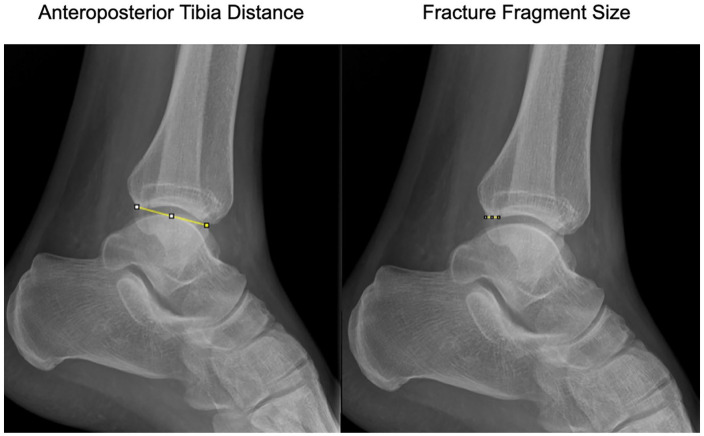
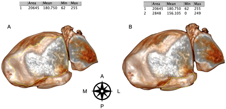
Methods: Morphology of the PMF was categorized initially by the CT scan as classified by Mason and Molloy on 180 trimalleolar ankle fractures. PM fracture fragment size on the lateral radiograph were compared to their respective 3D surface rendering CT reconstructions, by two independent observers. Morphology of the PMF was assessed using all preoperative radiographs as compared to 3D SR CT.
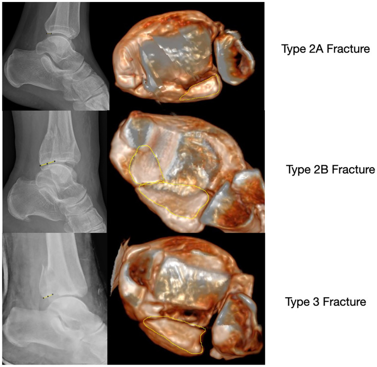
Results: On comparison of fracture fragment morphology, all fractures had poor categorization by plain radiographs although rotational pilon fractures (type 2A and 2B fractures) had the worse sensitivity and specificity (below 33% and below 50%, respectively). Radiographs underestimated joint involvement in type 2B fracture patterns because of the underappreciation of the posteromedial fragment.
Conclusion: This study shows that the use of plain radiographs to categorize morphology of PMFs is poor. The study adds to the ever-growing body of evidence on the inaccuracy of using plain radiographs in PMFs to plan treatment. Additional CT imaging is imperative to allow for appropriate treatment planning in the management of PMF. Smaller fracture fragments are more susceptible to inaccuracies, especially the rotational pilon subtypes (2A and 2B) because of the obliquity of the posteromedial fragment to the plane of the X-ray source.
Level of evidence: Level III, retrospective comparative study.
背景:本研究的目的是比较平面侧位x线片和3D表面渲染(SR) CT成像,使用Mason和Molloy分类来表征后踝骨折(PMF)的形态。原假设是x线平片和3D SR CT在形态学特征上没有差异。方法:采用Mason和Molloy对180例三踝踝关节骨折进行CT扫描,初步对PMF形态进行分类。两名独立观察员将侧位片上PM骨折碎片大小与各自的3D表面渲染CT重建进行比较。与3D SR CT相比,术前所有x线片评估PMF的形态。结果:对比骨折碎片形态,x线平片对所有骨折的分类都较差,但旋转皮隆骨折(2A型和2B型)的敏感性和特异性较差(分别低于33%和50%)。在2B型骨折中,由于对后内侧碎片的评价不足,x线片低估了关节受累程度。结论:利用x线平片对PMFs形态进行分类效果不佳。这项研究增加了越来越多的证据,证明在PMFs中使用x光平片来计划治疗是不准确的。额外的CT成像是必要的,以便在PMF的管理中制定适当的治疗计划。较小的骨折碎片更容易受到不准确的影响,特别是旋转pilon亚型(2A和2B),因为后内侧碎片与x射线源平面倾斜。证据等级:III级,回顾性比较研究。

 求助内容:
求助内容: 应助结果提醒方式:


