Bioactive scaffolds integrated with micro-precise spatiotemporal delivery and in vivo degradation tracking for complex tissue regeneration
Q1 Medicine
引用次数: 0
Abstract
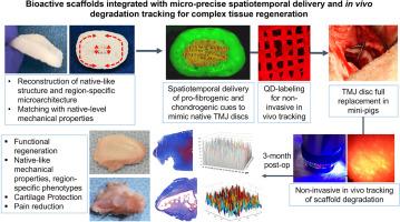
Three-dimensional (3D) printing has evolved to incorporate controlled delivery systems to guide the regeneration of complex tissues, with limited clinical translation. The challenges include the limited precision in spatiotemporal delivery and poorly understood in vivo scaffold degradation rates. Here, we report auspicious preclinical outcomes in the functional regeneration of temporomandibular joint (TMJ) discs of mini-pigs. TMJ disc has been an extremely challenging target for regenerative engineering given the uniquely heterogeneous matrix distribution and region-variant anisotropic orientation. We optimally implemented advanced 3D printing technologies with micro-precise spatiotemporal delivery to build anatomically correct, bioactive scaffolds with native-like regionally variant microstructure and mechanical properties. We also applied quantum dots (QDs) labeling of scaffolds to enable non-invasive in vivo degradation tracking. In mini-pigs, the scaffold implantation upon discectomy has successfully led to in-situ regeneration of TMJ discs by 3 months, exhibiting native-like heterogeneity and multi-scale mechanical properties without any sign of cartilage damage. In addition, our non-invasive imaging resulted in reliable in vivo tracking of scaffold degradation, exhibiting notably different degradation rates between individual animals. Our findings suggest a significant translational potential of our cell-free, bioactive scaffolds equipped with non-invasive tracking modality for in-situ tissue engineering of TMJ discs.

生物活性支架集成微精确时空传递和体内降解跟踪复杂组织再生
三维(3D)打印已经发展到结合控制输送系统来指导复杂组织的再生,但临床翻译有限。挑战包括有限的时空递送精度和对体内支架降解率的了解不足。在这里,我们报告了迷你猪颞下颌关节(TMJ)椎间盘功能再生的临床前结果。由于其独特的非均质矩阵分布和区域各向异性取向,TMJ椎间盘一直是再生工程中极具挑战性的目标。我们优化了先进的3D打印技术,通过微精确的时空传递来构建解剖学上正确的、具有生物活性的支架,这些支架具有类似于天然区域变化的微观结构和机械性能。我们还应用量子点(QDs)标记支架来实现无创体内降解跟踪。在小型猪中,椎间盘切除术后支架植入成功地导致TMJ椎间盘原位再生3个月,表现出类似天然的异质性和多尺度力学性能,没有任何软骨损伤的迹象。此外,我们的非侵入性成像导致了可靠的支架降解的体内跟踪,显示出个体动物之间明显不同的降解率。我们的研究结果表明,我们的无细胞、生物活性支架具有非侵入性跟踪模式,在TMJ椎间盘原位组织工程中具有重要的转化潜力。
本文章由计算机程序翻译,如有差异,请以英文原文为准。
求助全文
约1分钟内获得全文
求助全文
来源期刊

Engineered regeneration
Biomaterials, Medicine and Dentistry (General), Biotechnology, Biomedical Engineering
CiteScore
22.90
自引率
0.00%
发文量
0
审稿时长
33 days
 求助内容:
求助内容: 应助结果提醒方式:


