Camilla Toft Nielsen, Marius Henriksen, Cecilie Laubjerg Daugaard, Janus Uhd Nybing, Philip Hansen, Felix Müller, Henning Bliddal, Mikael Boesen, Henrik Gudbergsen
{"title":"The association between articular calcium crystal deposition and knee osteoarthritis, joint pain and inflammation: a cross-sectional study.","authors":"Camilla Toft Nielsen, Marius Henriksen, Cecilie Laubjerg Daugaard, Janus Uhd Nybing, Philip Hansen, Felix Müller, Henning Bliddal, Mikael Boesen, Henrik Gudbergsen","doi":"10.1007/s00256-025-04904-7","DOIUrl":null,"url":null,"abstract":"<p><strong>Objective: </strong>To explore in a cross-sectional fashion if overweight individuals with knee osteoarthritis (OA) and intraarticular calcium crystal (CaC) deposits experience more knee joint inflammation and knee pain compared with individuals without CaC deposits.</p><p><strong>Subjects and methods: </strong>We used pre-randomization imaging data from an RCT, the LOSE-IT trial. Participants with knee OA (clinical diagnosis of knee OA and KLG 1-3) had CT and 3 T MRI of the index knee. CaCs were assessed on CT using the Boston University Calcium Knee Score (BUCKS). The pain subscale of the Knee Injury and Osteoarthritis Outcome Score (KOOS) was used to assess knee pain and to estimate joint inflammation we used static and dynamic contrast-enhanced (DCE) MRI. An independent sample t-test was used to test for a significant difference in KOOS-pain and Analysis of Covariance (ANCOVA) models to test for differences in the static and DCE-MRI variables between the two groups.</p><p><strong>Results: </strong>Of the 158 participants with KOOS-pain available, 19 (12%) had CaC deposits, and of the 115 participants with MRI available, 13 (11.3%) had CaC deposits. We did not find a significant difference in mean KOOS-pain between the two groups; the mean difference was - 2.2 points (95%CI, - 10.86, 6.45). None of the MRI variables were associated with the presence of CaC deposits. Between-group differences were small for all MRI variables, with standardized mean differences ranging from small to medium (0.31-0.56).</p><p><strong>Conclusion: </strong>In individuals with knee OA, we did not find an association between intraarticular CaC deposits and an increase in knee joint inflammation or knee pain.</p>","PeriodicalId":21783,"journal":{"name":"Skeletal Radiology","volume":" ","pages":"1939-1947"},"PeriodicalIF":2.2000,"publicationDate":"2025-09-01","publicationTypes":"Journal Article","fieldsOfStudy":null,"isOpenAccess":false,"openAccessPdf":"https://www.ncbi.nlm.nih.gov/pmc/articles/PMC12241293/pdf/","citationCount":"0","resultStr":null,"platform":"Semanticscholar","paperid":null,"PeriodicalName":"Skeletal Radiology","FirstCategoryId":"3","ListUrlMain":"https://doi.org/10.1007/s00256-025-04904-7","RegionNum":3,"RegionCategory":"医学","ArticlePicture":[],"TitleCN":null,"AbstractTextCN":null,"PMCID":null,"EPubDate":"2025/3/14 0:00:00","PubModel":"Epub","JCR":"Q2","JCRName":"ORTHOPEDICS","Score":null,"Total":0}
引用次数: 0
Abstract
Objective: To explore in a cross-sectional fashion if overweight individuals with knee osteoarthritis (OA) and intraarticular calcium crystal (CaC) deposits experience more knee joint inflammation and knee pain compared with individuals without CaC deposits.
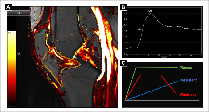
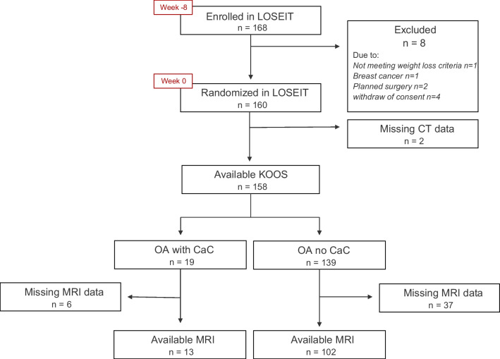
Subjects and methods: We used pre-randomization imaging data from an RCT, the LOSE-IT trial. Participants with knee OA (clinical diagnosis of knee OA and KLG 1-3) had CT and 3 T MRI of the index knee. CaCs were assessed on CT using the Boston University Calcium Knee Score (BUCKS). The pain subscale of the Knee Injury and Osteoarthritis Outcome Score (KOOS) was used to assess knee pain and to estimate joint inflammation we used static and dynamic contrast-enhanced (DCE) MRI. An independent sample t-test was used to test for a significant difference in KOOS-pain and Analysis of Covariance (ANCOVA) models to test for differences in the static and DCE-MRI variables between the two groups.
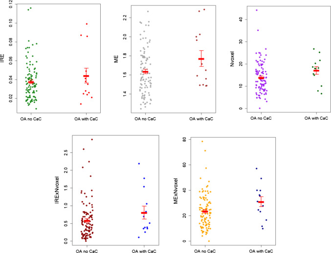
Results: Of the 158 participants with KOOS-pain available, 19 (12%) had CaC deposits, and of the 115 participants with MRI available, 13 (11.3%) had CaC deposits. We did not find a significant difference in mean KOOS-pain between the two groups; the mean difference was - 2.2 points (95%CI, - 10.86, 6.45). None of the MRI variables were associated with the presence of CaC deposits. Between-group differences were small for all MRI variables, with standardized mean differences ranging from small to medium (0.31-0.56).
Conclusion: In individuals with knee OA, we did not find an association between intraarticular CaC deposits and an increase in knee joint inflammation or knee pain.
期刊介绍:
Skeletal Radiology provides a forum for the dissemination of current knowledge and information dealing with disorders of the musculoskeletal system including the spine. While emphasizing the radiological aspects of the many varied skeletal abnormalities, the journal also adopts an interdisciplinary approach, reflecting the membership of the International Skeletal Society. Thus, the anatomical, pathological, physiological, clinical, metabolic and epidemiological aspects of the many entities affecting the skeleton receive appropriate consideration.
This is the Journal of the International Skeletal Society and the Official Journal of the Society of Skeletal Radiology and the Australasian Musculoskelelal Imaging Group.

 求助内容:
求助内容: 应助结果提醒方式:


