André Vaz, Kevin Rafael De Paula Morales, Eduardo Kaiser Ururahy Nunes Fonseca, Juliana Pato Serra Souza, Maria Júlia Silveira Rahal, Ludmila Mintzu Young, Leticia Muniz Pereira, Luiz Raphael Pereira Donoso Scoppetta, José Rodrigues Parga Filho
{"title":"Ring-like late gadolinium enhancement: differential diagnosis and mimics.","authors":"André Vaz, Kevin Rafael De Paula Morales, Eduardo Kaiser Ururahy Nunes Fonseca, Juliana Pato Serra Souza, Maria Júlia Silveira Rahal, Ludmila Mintzu Young, Leticia Muniz Pereira, Luiz Raphael Pereira Donoso Scoppetta, José Rodrigues Parga Filho","doi":"10.1590/0100-3984.2024.0111","DOIUrl":null,"url":null,"abstract":"<p><p>Advances in cardiac magnetic resonance have promoted tissue characterization with high spatial and contrast resolution, and late gadolinium enhancement (LGE) sequences have improved the detection of myocardial fibrosis. The distribution pattern of LGE facilitates differentiation between ischemic and nonischemic etiologies and aids in refining diagnoses within nonischemic cardiomyopathies, suggesting specific etiological factors. A distinctive nonischemic LGE pattern that has recently gained prominence is the ring-like pattern, defined as a subepicardial or mid-wall circumferential or semi-circumferential enhancement, which involves at least three contiguous segments within the same short-axis slice. Initially identified as a diagnostic marker for desmoplakin and filamin C-related cardiomyopathies, the pattern has been reported in nongenetic conditions; nevertheless, it remains an uncommon finding in these diseases. In this article, we aim to present the differential diagnoses of ring-like LGE and its mimics. The combination of epidemiological, clinical, electrocardiographic, and additional features enables a focused refinement of the differential diagnosis associated with ring-like LGE.</p>","PeriodicalId":20842,"journal":{"name":"Radiologia Brasileira","volume":"58 ","pages":"e20240111"},"PeriodicalIF":0.0000,"publicationDate":"2025-02-27","publicationTypes":"Journal Article","fieldsOfStudy":null,"isOpenAccess":false,"openAccessPdf":"https://www.ncbi.nlm.nih.gov/pmc/articles/PMC11905226/pdf/","citationCount":"0","resultStr":null,"platform":"Semanticscholar","paperid":null,"PeriodicalName":"Radiologia Brasileira","FirstCategoryId":"1085","ListUrlMain":"https://doi.org/10.1590/0100-3984.2024.0111","RegionNum":0,"RegionCategory":null,"ArticlePicture":[],"TitleCN":null,"AbstractTextCN":null,"PMCID":null,"EPubDate":"2025/1/1 0:00:00","PubModel":"eCollection","JCR":"Q3","JCRName":"Medicine","Score":null,"Total":0}
引用次数: 0
Abstract
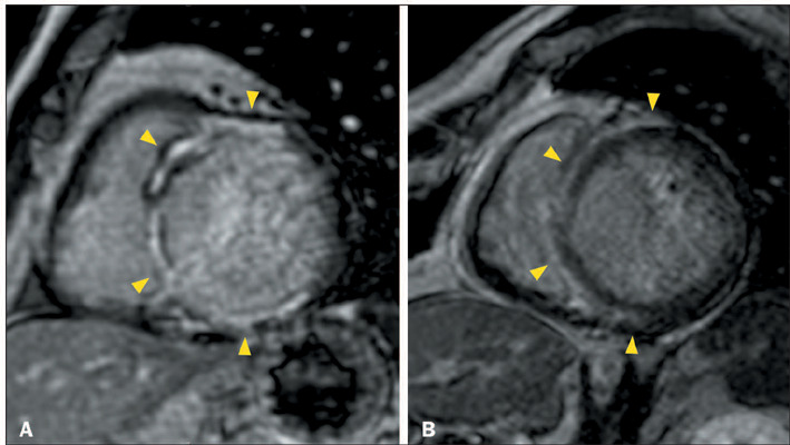
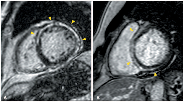
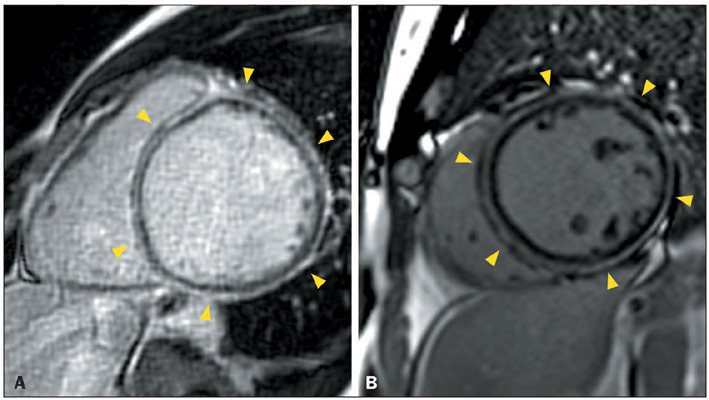
Advances in cardiac magnetic resonance have promoted tissue characterization with high spatial and contrast resolution, and late gadolinium enhancement (LGE) sequences have improved the detection of myocardial fibrosis. The distribution pattern of LGE facilitates differentiation between ischemic and nonischemic etiologies and aids in refining diagnoses within nonischemic cardiomyopathies, suggesting specific etiological factors. A distinctive nonischemic LGE pattern that has recently gained prominence is the ring-like pattern, defined as a subepicardial or mid-wall circumferential or semi-circumferential enhancement, which involves at least three contiguous segments within the same short-axis slice. Initially identified as a diagnostic marker for desmoplakin and filamin C-related cardiomyopathies, the pattern has been reported in nongenetic conditions; nevertheless, it remains an uncommon finding in these diseases. In this article, we aim to present the differential diagnoses of ring-like LGE and its mimics. The combination of epidemiological, clinical, electrocardiographic, and additional features enables a focused refinement of the differential diagnosis associated with ring-like LGE.

 求助内容:
求助内容: 应助结果提醒方式:


