Adib Al-Haj Husain, Victor Mergen, Silvio Valdec, Nadin Al-Haj Husain, Bernd Stadlinger, Harald Essig, Thomas Frauenfelder, Peter Kessler, Suen An Nynke Lie, Hatem Alkadhi, Sebastian Winklhofer
{"title":"Comparison of cone-beam computed tomography with photon-counting detector computed tomography for dental implant surgery.","authors":"Adib Al-Haj Husain, Victor Mergen, Silvio Valdec, Nadin Al-Haj Husain, Bernd Stadlinger, Harald Essig, Thomas Frauenfelder, Peter Kessler, Suen An Nynke Lie, Hatem Alkadhi, Sebastian Winklhofer","doi":"10.1186/s40729-025-00611-z","DOIUrl":null,"url":null,"abstract":"<p><strong>Purpose: </strong>To compare cone-beam computed tomography (CBCT) with photon-counting detector computed tomography (PCD-CT) at equivalent radiation doses, focusing on qualitative and quantitative parameters relevant to dental implant surgery.</p><p><strong>Methods: </strong>This ex vivo comparative study of porcine specimens assessed five imaging protocols with both CBCT and PCD-CT at three effective radiation dose levels (high: 360µSv, standard: 145µSv, low: 20µSv) to evaluate image quality, artifact burden, metal artifact susceptibility, and quantitative bone measurements in the mandibular region. Three blinded readers analyzed the data using a 5-point Likert scale (5 = highest to 1 = lowest rating) and performed linear bone measurements at implant planning sites. Statistical analysis included descriptive statistics and inter-reader reliability assessment using intraclass correlation coefficients (ICC).</p><p><strong>Results: </strong>Each reader evaluated 30 data sets (12 CBCT, 18 PCD-CT), with 24 implant planning sites per imaging protocol. High-dose PCD-CT demonstrated the best image quality and diagnostic interpretability (4.89 ± 0.27), followed by standard-dose PCD-CT and CBCT (4.50 ± 0.73; 4.33 ± 0.61), with low-dose protocols showing intermediate quality with higher artifact burden. In comparison to CBCT, PCD-CT demonstrated superior performance in reducing implant-induced artifacts across all protocols. Quantitative bone measurements showed minimal variability, meeting clinical precision requirements for computer-assisted implant surgery. Both qualitative (ICCs:0.70-0.89; p < 0.001) and quantitative (ICCs:0.79-1; p < 0.001) analyses demonstrated high reliability, regardless of the reader's experience.</p><p><strong>Conclusions: </strong>PCD-CT demonstrated superior image quality and reduced artifacts compared with CBCT at all radiation dose levels. These findings highlight PCD-CT's potential to enhance implant planning and improve clinical outcomes with reduced radiation exposure while maintaining diagnostic accuracy.</p>","PeriodicalId":14076,"journal":{"name":"International Journal of Implant Dentistry","volume":"11 1","pages":"21"},"PeriodicalIF":4.0000,"publicationDate":"2025-03-13","publicationTypes":"Journal Article","fieldsOfStudy":null,"isOpenAccess":false,"openAccessPdf":"https://www.ncbi.nlm.nih.gov/pmc/articles/PMC11906956/pdf/","citationCount":"0","resultStr":null,"platform":"Semanticscholar","paperid":null,"PeriodicalName":"International Journal of Implant Dentistry","FirstCategoryId":"3","ListUrlMain":"https://doi.org/10.1186/s40729-025-00611-z","RegionNum":3,"RegionCategory":"医学","ArticlePicture":[],"TitleCN":null,"AbstractTextCN":null,"PMCID":null,"EPubDate":"","PubModel":"","JCR":"Q1","JCRName":"DENTISTRY, ORAL SURGERY & MEDICINE","Score":null,"Total":0}
引用次数: 0
Abstract
Purpose: To compare cone-beam computed tomography (CBCT) with photon-counting detector computed tomography (PCD-CT) at equivalent radiation doses, focusing on qualitative and quantitative parameters relevant to dental implant surgery.
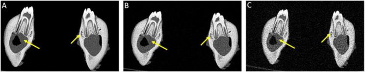
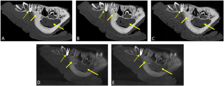
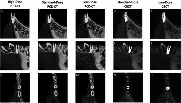
Methods: This ex vivo comparative study of porcine specimens assessed five imaging protocols with both CBCT and PCD-CT at three effective radiation dose levels (high: 360µSv, standard: 145µSv, low: 20µSv) to evaluate image quality, artifact burden, metal artifact susceptibility, and quantitative bone measurements in the mandibular region. Three blinded readers analyzed the data using a 5-point Likert scale (5 = highest to 1 = lowest rating) and performed linear bone measurements at implant planning sites. Statistical analysis included descriptive statistics and inter-reader reliability assessment using intraclass correlation coefficients (ICC).
Results: Each reader evaluated 30 data sets (12 CBCT, 18 PCD-CT), with 24 implant planning sites per imaging protocol. High-dose PCD-CT demonstrated the best image quality and diagnostic interpretability (4.89 ± 0.27), followed by standard-dose PCD-CT and CBCT (4.50 ± 0.73; 4.33 ± 0.61), with low-dose protocols showing intermediate quality with higher artifact burden. In comparison to CBCT, PCD-CT demonstrated superior performance in reducing implant-induced artifacts across all protocols. Quantitative bone measurements showed minimal variability, meeting clinical precision requirements for computer-assisted implant surgery. Both qualitative (ICCs:0.70-0.89; p < 0.001) and quantitative (ICCs:0.79-1; p < 0.001) analyses demonstrated high reliability, regardless of the reader's experience.
Conclusions: PCD-CT demonstrated superior image quality and reduced artifacts compared with CBCT at all radiation dose levels. These findings highlight PCD-CT's potential to enhance implant planning and improve clinical outcomes with reduced radiation exposure while maintaining diagnostic accuracy.
期刊介绍:
The International Journal of Implant Dentistry is a peer-reviewed open access journal published under the SpringerOpen brand. The journal is dedicated to promoting the exchange and discussion of all research areas relevant to implant dentistry in the form of systematic literature or invited reviews, prospective and retrospective clinical studies, clinical case reports, basic laboratory and animal research, and articles on material research and engineering.
 求助内容:
求助内容: 应助结果提醒方式:


