Comparison of ProExC and p16ink4a Biological Markers in Lesional Smears With the Immunocytochemical Method and Relationship With Human Papillomavirus in Liquid-based Cervicovaginal Specimens.
{"title":"Comparison of ProExC and p16ink4a Biological Markers in Lesional Smears With the Immunocytochemical Method and Relationship With Human Papillomavirus in Liquid-based Cervicovaginal Specimens.","authors":"Zeynep Turkmen Usta, Zeynep Sagnak Yilmaz, Safak Ersoz, Sevdegul Aydin Mungan, Umit Cobanoglu, Suleyman Guven","doi":"10.4103/joc.joc_57_24","DOIUrl":null,"url":null,"abstract":"<p><strong>Background: </strong>This study investigated the determination of human papillomavirus (HPV) types in smears with and without lesions, and the sensitivity, specificity, positive predictive value, and negative predictive value of the ProExC and p16 biomarkers in smears with lesions.</p><p><strong>Materials and methods: </strong>A total of 192 cervicovaginal smears were included in the study. ProExC (BD) and p16ink4a antibodies were applied to the lesion-containing samples by immunocytochemical method. If HPV was present, its type was determined. Patient biopsy specimens were used as a gold standard to confirm the lesion type. In addition, atypical squamous cells of undetermined significance, low-grade squamous intraepithelial lesion (SIL), atypical squamous cells, cannot exclude high-grade SIL (HSIL), and HSIL, atypical glandular cells, and the relationship between two biomarkers in cases diagnosed with squamous cell carcinoma were investigated.</p><p><strong>Results: </strong>Of these, 192 cases included in our study, 119 had lesional smears and 73 had no lesional cells. Of the 191 cases in which HPV was investigated, 105 were negative and 86 were positive for HPV types 16, 18, and others. A statistically significant difference was found between HPV positivity and smears with lesions (<i>P</i> = 0.0001). p16 and ProExC positivity was extensive in cases with more severe lesions. A strong correlation was observed between high-risk HPV (+) and HSIL-detected cases.</p><p><strong>Conclusion: </strong>ProExC and p16 are biomarkers that facilitate the diagnosis of HSIL. Nuclear staining for the ProExC marker is easier to apply to cytological samples than p16.</p>","PeriodicalId":50217,"journal":{"name":"Journal of Cytology","volume":"42 1","pages":"20-26"},"PeriodicalIF":1.0000,"publicationDate":"2024-01-01","publicationTypes":"Journal Article","fieldsOfStudy":null,"isOpenAccess":false,"openAccessPdf":"https://www.ncbi.nlm.nih.gov/pmc/articles/PMC11896117/pdf/","citationCount":"0","resultStr":null,"platform":"Semanticscholar","paperid":null,"PeriodicalName":"Journal of Cytology","FirstCategoryId":"3","ListUrlMain":"https://doi.org/10.4103/joc.joc_57_24","RegionNum":4,"RegionCategory":"医学","ArticlePicture":[],"TitleCN":null,"AbstractTextCN":null,"PMCID":null,"EPubDate":"2025/2/11 0:00:00","PubModel":"Epub","JCR":"Q4","JCRName":"MEDICAL LABORATORY TECHNOLOGY","Score":null,"Total":0}
引用次数: 0
Abstract
Background: This study investigated the determination of human papillomavirus (HPV) types in smears with and without lesions, and the sensitivity, specificity, positive predictive value, and negative predictive value of the ProExC and p16 biomarkers in smears with lesions.
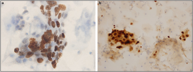
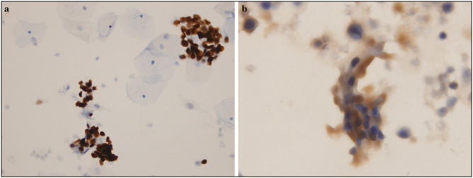
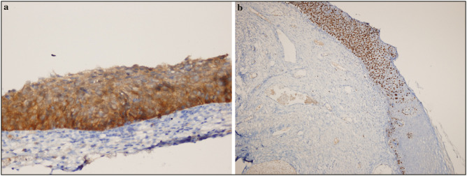
Materials and methods: A total of 192 cervicovaginal smears were included in the study. ProExC (BD) and p16ink4a antibodies were applied to the lesion-containing samples by immunocytochemical method. If HPV was present, its type was determined. Patient biopsy specimens were used as a gold standard to confirm the lesion type. In addition, atypical squamous cells of undetermined significance, low-grade squamous intraepithelial lesion (SIL), atypical squamous cells, cannot exclude high-grade SIL (HSIL), and HSIL, atypical glandular cells, and the relationship between two biomarkers in cases diagnosed with squamous cell carcinoma were investigated.
Results: Of these, 192 cases included in our study, 119 had lesional smears and 73 had no lesional cells. Of the 191 cases in which HPV was investigated, 105 were negative and 86 were positive for HPV types 16, 18, and others. A statistically significant difference was found between HPV positivity and smears with lesions (P = 0.0001). p16 and ProExC positivity was extensive in cases with more severe lesions. A strong correlation was observed between high-risk HPV (+) and HSIL-detected cases.
Conclusion: ProExC and p16 are biomarkers that facilitate the diagnosis of HSIL. Nuclear staining for the ProExC marker is easier to apply to cytological samples than p16.
期刊介绍:
The Journal of Cytology is the official Quarterly publication of the Indian Academy of Cytologists. It is in the 25th year of publication in the year 2008. The journal covers all aspects of diagnostic cytology, including fine needle aspiration cytology, gynecological and non-gynecological cytology. Articles on ancillary techniques, like cytochemistry, immunocytochemistry, electron microscopy, molecular cytopathology, as applied to cytological material are also welcome. The journal gives preference to clinically oriented studies over experimental and animal studies. The Journal would publish peer-reviewed original research papers, case reports, systematic reviews, meta-analysis, and debates.
 求助内容:
求助内容: 应助结果提醒方式:


