Dose- and sex-dependent effects on umbilical cord-derived mesenchymal stem cell efficacy in regeneration of a full-thickness tendon defect in a rat model.
{"title":"Dose- and sex-dependent effects on umbilical cord-derived mesenchymal stem cell efficacy in regeneration of a full-thickness tendon defect in a rat model.","authors":"Ji-Hye Yea, Chris Hyunchul Jo","doi":"10.5397/cise.2024.00626","DOIUrl":null,"url":null,"abstract":"<p><strong>Background: </strong>Mesenchymal stem cells (MSCs) have shown potential in regenerative medicine. In the present study the effects of MSC dosage and recipient sex on tendon regeneration were evaluated.</p><p><strong>Methods: </strong>A full-thickness tendon defect (FTTD) was created on supraspinatus tendons (SSTs) of rats and cryoprotective solution (CPS) and MSCs (0.05, 0.1 and 0.5 million MSCs [M-MSC] for female groups and 1.0 M-MSC for both female and male groups) were applied. After 2 and 4 weeks, macroscopic and histological evaluations were performed.</p><p><strong>Results: </strong>Total macroscopic scores were improved in all MSC groups compared with the CPS group, with no significant differences among the MSC groups. Furthermore, all MSC groups had lower total degenerative scores than the CPS group; however, only 0.1 M-MSC, 0.5 M-MSC, and 1 M-MSC groups showed significantly improved hyalinization compared with the CPS group at 4 weeks. Collagen organization and coherence were higher in all MSC groups than in the CPS group at both 2 and 4 weeks; however, 0.5 M-MSC and 1 M-MSC groups scored better than the 0.05 M-MSC group at 4 weeks. Heterotopic matrix analysis revealed smaller glycosaminoglycan (GAG)-rich areas in the 0.1 M-MSC, 0.5 M-MSC, and 1 M-MSC groups compared with the CPS group at 4 weeks. Overall, macroscopic and histological evaluations were not significantly different between female and male groups except for GAG-rich area.</p><p><strong>Conclusions: </strong>The MSC dosage affected collagen and heterotopic matrix formation in a FTTD rat model; however, the efficacy of MSCs (1.0 M dose) in collagen regeneration was not affected based on the sex of the recipient. Level of evidence: I.</p>","PeriodicalId":33981,"journal":{"name":"Clinics in Shoulder and Elbow","volume":"28 1","pages":"49-59"},"PeriodicalIF":1.7000,"publicationDate":"2025-03-01","publicationTypes":"Journal Article","fieldsOfStudy":null,"isOpenAccess":false,"openAccessPdf":"https://www.ncbi.nlm.nih.gov/pmc/articles/PMC11938924/pdf/","citationCount":"0","resultStr":null,"platform":"Semanticscholar","paperid":null,"PeriodicalName":"Clinics in Shoulder and Elbow","FirstCategoryId":"1085","ListUrlMain":"https://doi.org/10.5397/cise.2024.00626","RegionNum":0,"RegionCategory":null,"ArticlePicture":[],"TitleCN":null,"AbstractTextCN":null,"PMCID":null,"EPubDate":"2025/2/17 0:00:00","PubModel":"Epub","JCR":"Q2","JCRName":"ORTHOPEDICS","Score":null,"Total":0}
引用次数: 0
Abstract
Background: Mesenchymal stem cells (MSCs) have shown potential in regenerative medicine. In the present study the effects of MSC dosage and recipient sex on tendon regeneration were evaluated.
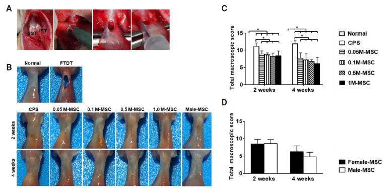
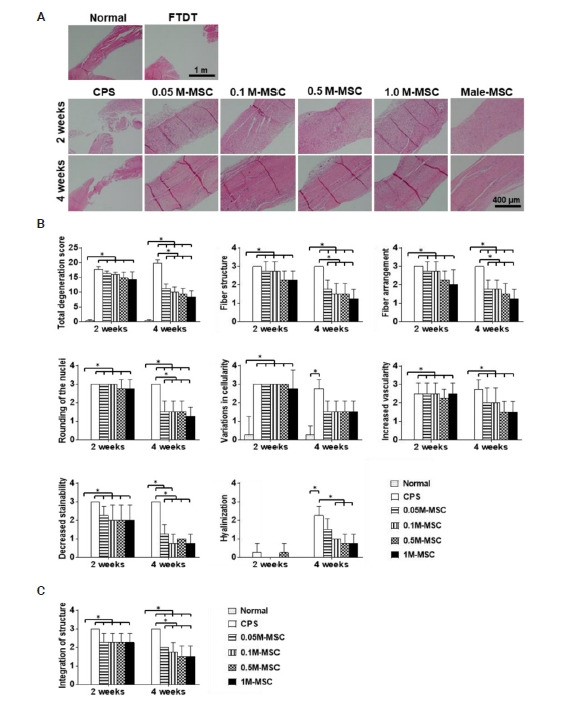
Methods: A full-thickness tendon defect (FTTD) was created on supraspinatus tendons (SSTs) of rats and cryoprotective solution (CPS) and MSCs (0.05, 0.1 and 0.5 million MSCs [M-MSC] for female groups and 1.0 M-MSC for both female and male groups) were applied. After 2 and 4 weeks, macroscopic and histological evaluations were performed.
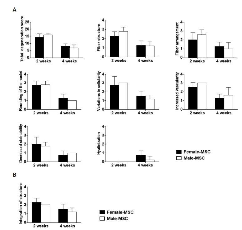
Results: Total macroscopic scores were improved in all MSC groups compared with the CPS group, with no significant differences among the MSC groups. Furthermore, all MSC groups had lower total degenerative scores than the CPS group; however, only 0.1 M-MSC, 0.5 M-MSC, and 1 M-MSC groups showed significantly improved hyalinization compared with the CPS group at 4 weeks. Collagen organization and coherence were higher in all MSC groups than in the CPS group at both 2 and 4 weeks; however, 0.5 M-MSC and 1 M-MSC groups scored better than the 0.05 M-MSC group at 4 weeks. Heterotopic matrix analysis revealed smaller glycosaminoglycan (GAG)-rich areas in the 0.1 M-MSC, 0.5 M-MSC, and 1 M-MSC groups compared with the CPS group at 4 weeks. Overall, macroscopic and histological evaluations were not significantly different between female and male groups except for GAG-rich area.
Conclusions: The MSC dosage affected collagen and heterotopic matrix formation in a FTTD rat model; however, the efficacy of MSCs (1.0 M dose) in collagen regeneration was not affected based on the sex of the recipient. Level of evidence: I.

 求助内容:
求助内容: 应助结果提醒方式:


