E P Yanysheva, P A Melnikov, D A Chudakova, M V Shirmanova, V P Baklaushev, G M Yusubalieva
{"title":"Migration of Regulatory T Cells to the Peritumor Microenvironment of Experimental Glioblastoma.","authors":"E P Yanysheva, P A Melnikov, D A Chudakova, M V Shirmanova, V P Baklaushev, G M Yusubalieva","doi":"10.17691/stm2025.17.1.07","DOIUrl":null,"url":null,"abstract":"<p><p>Glioblastoma is the most aggressive primary brain tumor with poor prognosis characterized by resistance to standard treatments and immune evasion. Regulatory T lymphocytes (Tregs) play a key role in immune suppression in the tumor microenvironment and can be used as targets for malignant gliomas therapy. <b>The aim of the investigation</b> is to study migration of Tregs to the tumor site in the process of dynamic glioblastoma growth on the transgenic C57Bl/6-FoxP3-eGFP mouse line.</p><p><strong>Materials and methods: </strong>The study was performed using the C57Bl/6-FoxP3-eGFP mouse strain, which allows for the detection of FoxP3-positive Tregs by fluorescent signal. Orthotopic glioblastomas were implanted by stereotactic injection of fluorescently labeled GL-261-BFP and GL-261-mScarlet tumor cell lines. Intravital confocal microscopy was used to monitor infiltration of the tumor site by immune cells, visualized by intravenous injection of fluorescently labeled antibodies against CD45. The results of intravital microscopy were confirmed by histological and immunohistochemical examination on days 3, 6, 9, 14, and 16 after the implantation. To assess the immunological status, tumor-infiltrating lymphocytes (TILs) were isolated from the brain and Tregs were counted using a flow cytometer (immediately after isolation and after cultivation for 2 weeks).</p><p><strong>Results: </strong>Intravital microscopy and brain slice studies have demonstrated infiltration of the glioblastoma site by Tregs, with the proportion of Tregs increasing with tumor progression (the increase in the absolute number of Treg was proportional to the increase in the number of glioma cells). Subsequent co-cultivation of isolated TILs with glioma cells revealed increase of Treg population within 2 weeks from 2.8% to >40%, confirming the activating effect of glioblastoma with respect to Tregs.</p><p><strong>Conclusion: </strong>The dynamics of GL-261 glioma microenvironment infiltration by Tregs has been investigated. The glioblastoma cells were shown to activate Tregs in the peritumor space <i>in vivo</i> and to promote their selective expansion when co-cultured with TILs <i>in vitro</i>. These data can be used for further studies on C57Bl/6-FoxP3-eGFP mice to find approaches to inactivate Tregs in glioblastoma.</p>","PeriodicalId":520289,"journal":{"name":"Sovremennye tekhnologii v meditsine","volume":"17 1","pages":"70-78"},"PeriodicalIF":0.0000,"publicationDate":"2025-01-01","publicationTypes":"Journal Article","fieldsOfStudy":null,"isOpenAccess":false,"openAccessPdf":"https://www.ncbi.nlm.nih.gov/pmc/articles/PMC11892569/pdf/","citationCount":"0","resultStr":null,"platform":"Semanticscholar","paperid":null,"PeriodicalName":"Sovremennye tekhnologii v meditsine","FirstCategoryId":"1085","ListUrlMain":"https://doi.org/10.17691/stm2025.17.1.07","RegionNum":0,"RegionCategory":null,"ArticlePicture":[],"TitleCN":null,"AbstractTextCN":null,"PMCID":null,"EPubDate":"2025/2/28 0:00:00","PubModel":"Epub","JCR":"","JCRName":"","Score":null,"Total":0}
引用次数: 0
Abstract
Glioblastoma is the most aggressive primary brain tumor with poor prognosis characterized by resistance to standard treatments and immune evasion. Regulatory T lymphocytes (Tregs) play a key role in immune suppression in the tumor microenvironment and can be used as targets for malignant gliomas therapy. The aim of the investigation is to study migration of Tregs to the tumor site in the process of dynamic glioblastoma growth on the transgenic C57Bl/6-FoxP3-eGFP mouse line.
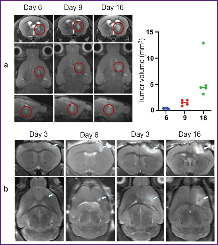
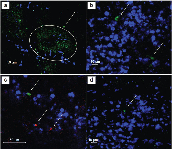
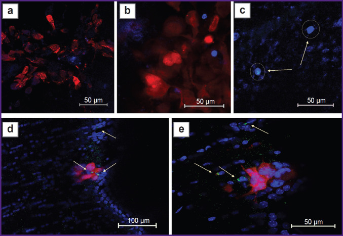
Materials and methods: The study was performed using the C57Bl/6-FoxP3-eGFP mouse strain, which allows for the detection of FoxP3-positive Tregs by fluorescent signal. Orthotopic glioblastomas were implanted by stereotactic injection of fluorescently labeled GL-261-BFP and GL-261-mScarlet tumor cell lines. Intravital confocal microscopy was used to monitor infiltration of the tumor site by immune cells, visualized by intravenous injection of fluorescently labeled antibodies against CD45. The results of intravital microscopy were confirmed by histological and immunohistochemical examination on days 3, 6, 9, 14, and 16 after the implantation. To assess the immunological status, tumor-infiltrating lymphocytes (TILs) were isolated from the brain and Tregs were counted using a flow cytometer (immediately after isolation and after cultivation for 2 weeks).
Results: Intravital microscopy and brain slice studies have demonstrated infiltration of the glioblastoma site by Tregs, with the proportion of Tregs increasing with tumor progression (the increase in the absolute number of Treg was proportional to the increase in the number of glioma cells). Subsequent co-cultivation of isolated TILs with glioma cells revealed increase of Treg population within 2 weeks from 2.8% to >40%, confirming the activating effect of glioblastoma with respect to Tregs.
Conclusion: The dynamics of GL-261 glioma microenvironment infiltration by Tregs has been investigated. The glioblastoma cells were shown to activate Tregs in the peritumor space in vivo and to promote their selective expansion when co-cultured with TILs in vitro. These data can be used for further studies on C57Bl/6-FoxP3-eGFP mice to find approaches to inactivate Tregs in glioblastoma.

 求助内容:
求助内容: 应助结果提醒方式:


