Sevde Nur Emir, Merve Gürsu, Safiye Sanem Dereli Bulut
{"title":"Evaluating the potential of abbreviated MRI protocols for liver metastasis detection: a study in colorectal cancer patients.","authors":"Sevde Nur Emir, Merve Gürsu, Safiye Sanem Dereli Bulut","doi":"10.5114/pjr/196906","DOIUrl":null,"url":null,"abstract":"<p><strong>Purpose: </strong>To evaluate the diagnostic accuracy of different abbreviated magnetic resonance imaging (AMRI) protocols consisting of dynamic enhanced + T2-weighted imaging (T2W) and diffusion-weighted imaging (DWI) + T2W for the detection and characterization of liver metastases in a patient group with known colorectal cancer.</p><p><strong>Material and methods: </strong>A total of 197 hepatic lesions were retrospectively analyzed across 3 different MRI sets: AMRI-1 (dynamic enhanced + T2W), AMRI-2 (DWI + T2W), and a standard MRI protocol. The patient cohort included 100 individuals (63 males, 37 females) with a mean age of 62.6 years (SD: 11.1 years). Lesions were characterized as benign, malignant, or indeterminate based on histopathology, positron emission tomography-computed tomography (PET-CT), and follow-up imaging.</p><p><strong>Results: </strong>The standard MRI protocol identified 197 liver lesions (175 metastatic, 18 benign, and 4 indeterminate); 142 lesions (72.1%) were larger than 10 mm, with the majority being metastatic (140/142). Radiologist 1 identified 195 lesions using the AMRI-1 protocol (175 metastatic, 15 benign, and 5 indeterminate). The sensitivity per lesion was 89.7% (95% CI: 0.85-0.93). Radiologist 2 identified 183 lesions using the AMRI-2 protocol (169 metastatic, 6 benign, and 8 indeterminate). The sensitivity per lesion was 92.3% (95% CI: 0.88-0.95). No statistically significant difference was found in sensitivity between the AMRI-1 and AMRI-2 and standard MRI protocols (<i>p</i> > 0.05).</p><p><strong>Conclusions: </strong>The standard MRI protocol demonstrated the highest sensitivity and specificity for detecting and characterizing liver metastases. However, differences between the protocols were not statistically significant. Abbreviated MRI protocols, particularly the AMRI-2 protocol incorporating diffusion-weighted imaging, could serve as an effective alternative for routine clinical practice.</p>","PeriodicalId":94174,"journal":{"name":"Polish journal of radiology","volume":"90 ","pages":"e19-e25"},"PeriodicalIF":0.0000,"publicationDate":"2025-01-15","publicationTypes":"Journal Article","fieldsOfStudy":null,"isOpenAccess":false,"openAccessPdf":"https://www.ncbi.nlm.nih.gov/pmc/articles/PMC11891548/pdf/","citationCount":"0","resultStr":null,"platform":"Semanticscholar","paperid":null,"PeriodicalName":"Polish journal of radiology","FirstCategoryId":"1085","ListUrlMain":"https://doi.org/10.5114/pjr/196906","RegionNum":0,"RegionCategory":null,"ArticlePicture":[],"TitleCN":null,"AbstractTextCN":null,"PMCID":null,"EPubDate":"2025/1/1 0:00:00","PubModel":"eCollection","JCR":"","JCRName":"","Score":null,"Total":0}
引用次数: 0
Abstract
Purpose: To evaluate the diagnostic accuracy of different abbreviated magnetic resonance imaging (AMRI) protocols consisting of dynamic enhanced + T2-weighted imaging (T2W) and diffusion-weighted imaging (DWI) + T2W for the detection and characterization of liver metastases in a patient group with known colorectal cancer.
Material and methods: A total of 197 hepatic lesions were retrospectively analyzed across 3 different MRI sets: AMRI-1 (dynamic enhanced + T2W), AMRI-2 (DWI + T2W), and a standard MRI protocol. The patient cohort included 100 individuals (63 males, 37 females) with a mean age of 62.6 years (SD: 11.1 years). Lesions were characterized as benign, malignant, or indeterminate based on histopathology, positron emission tomography-computed tomography (PET-CT), and follow-up imaging.
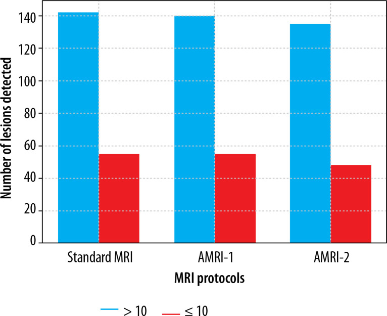
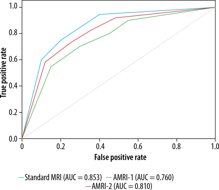
Results: The standard MRI protocol identified 197 liver lesions (175 metastatic, 18 benign, and 4 indeterminate); 142 lesions (72.1%) were larger than 10 mm, with the majority being metastatic (140/142). Radiologist 1 identified 195 lesions using the AMRI-1 protocol (175 metastatic, 15 benign, and 5 indeterminate). The sensitivity per lesion was 89.7% (95% CI: 0.85-0.93). Radiologist 2 identified 183 lesions using the AMRI-2 protocol (169 metastatic, 6 benign, and 8 indeterminate). The sensitivity per lesion was 92.3% (95% CI: 0.88-0.95). No statistically significant difference was found in sensitivity between the AMRI-1 and AMRI-2 and standard MRI protocols (p > 0.05).
Conclusions: The standard MRI protocol demonstrated the highest sensitivity and specificity for detecting and characterizing liver metastases. However, differences between the protocols were not statistically significant. Abbreviated MRI protocols, particularly the AMRI-2 protocol incorporating diffusion-weighted imaging, could serve as an effective alternative for routine clinical practice.

 求助内容:
求助内容: 应助结果提醒方式:


