Tatiana Blokhina, Tatiana Kirichenko, Yuliya Markina, Ulyana Khovantseva, Ivan Melnikov, Olga Guseva, Sergey Bazanovich, Sergey Kozlov, Alexander Orekhov
{"title":"Features of the monocyte inflammatory response in patients with premature coronary artery disease.","authors":"Tatiana Blokhina, Tatiana Kirichenko, Yuliya Markina, Ulyana Khovantseva, Ivan Melnikov, Olga Guseva, Sergey Bazanovich, Sergey Kozlov, Alexander Orekhov","doi":"10.52601/bpr.2024.240030","DOIUrl":null,"url":null,"abstract":"<p><p>The purpose of this study was to examine the secretion of inflammatory cytokines by cultured monocytes/macrophages in patients with premature coronary artery disease (CAD). The study included 38 patients with premature CAD and 35 patients without CAD. A primary culture of CD14+ monocytes was obtained by immunomagnetic separation. The inflammatory response was induced by incubation of a cell culture with lipopolysaccharide (LPS) for 24 hours on Days 1 and 6. Basal and LPS-stimulated secretion of the cytokines, tumor necrosis factor-α (TNF-α), interleukin-1β (IL-1β), interleukin-6 (IL-6), interleukin-8 (IL-8) and monocyte chemotactic protein-1 (MCP-1) was assessed by enzyme immunoassay on Days 2 and 7 of cultivation. The level of basal secretion of TNF-α, IL-1β, IL-6, MCP-1 was higher in patients with CAD compared to patients in the control group. The levels of re-stimulated TNF-α secretion and the levels of LPS-stimulated and re-stimulated IL-1β secretion on the second and sixth days were also higher in patients with CAD. LPS-stimulated MCP-1 secretion on the second day did not differ in patients of both groups, but re-stimulated MCP-1 secretion was higher in patients with CAD. The results of logistic regression analysis showed that the basal secretion levels of IL-1β and IL-6 were independently associated with premature CAD, along with smoking, body mass index and serum HDL-cholesterol levels.</p>","PeriodicalId":93906,"journal":{"name":"Biophysics reports","volume":"11 1","pages":"12-17"},"PeriodicalIF":0.0000,"publicationDate":"2025-02-28","publicationTypes":"Journal Article","fieldsOfStudy":null,"isOpenAccess":false,"openAccessPdf":"https://www.ncbi.nlm.nih.gov/pmc/articles/PMC11891073/pdf/","citationCount":"0","resultStr":null,"platform":"Semanticscholar","paperid":null,"PeriodicalName":"Biophysics reports","FirstCategoryId":"1085","ListUrlMain":"https://doi.org/10.52601/bpr.2024.240030","RegionNum":0,"RegionCategory":null,"ArticlePicture":[],"TitleCN":null,"AbstractTextCN":null,"PMCID":null,"EPubDate":"","PubModel":"","JCR":"","JCRName":"","Score":null,"Total":0}
引用次数: 0
Abstract
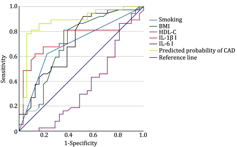
The purpose of this study was to examine the secretion of inflammatory cytokines by cultured monocytes/macrophages in patients with premature coronary artery disease (CAD). The study included 38 patients with premature CAD and 35 patients without CAD. A primary culture of CD14+ monocytes was obtained by immunomagnetic separation. The inflammatory response was induced by incubation of a cell culture with lipopolysaccharide (LPS) for 24 hours on Days 1 and 6. Basal and LPS-stimulated secretion of the cytokines, tumor necrosis factor-α (TNF-α), interleukin-1β (IL-1β), interleukin-6 (IL-6), interleukin-8 (IL-8) and monocyte chemotactic protein-1 (MCP-1) was assessed by enzyme immunoassay on Days 2 and 7 of cultivation. The level of basal secretion of TNF-α, IL-1β, IL-6, MCP-1 was higher in patients with CAD compared to patients in the control group. The levels of re-stimulated TNF-α secretion and the levels of LPS-stimulated and re-stimulated IL-1β secretion on the second and sixth days were also higher in patients with CAD. LPS-stimulated MCP-1 secretion on the second day did not differ in patients of both groups, but re-stimulated MCP-1 secretion was higher in patients with CAD. The results of logistic regression analysis showed that the basal secretion levels of IL-1β and IL-6 were independently associated with premature CAD, along with smoking, body mass index and serum HDL-cholesterol levels.

 求助内容:
求助内容: 应助结果提醒方式:


