Ahmed Altaf, Muhammad Sami Alam, Sibgha Khan, Ali Azan, Fatima Mubarak, Edmond Knopp, Khan Siddiqui, Syed Ather Enam
{"title":"Initial insights into post-contrast enhancement in ultra-low-field MRI: Case Report.","authors":"Ahmed Altaf, Muhammad Sami Alam, Sibgha Khan, Ali Azan, Fatima Mubarak, Edmond Knopp, Khan Siddiqui, Syed Ather Enam","doi":"10.3389/fnimg.2025.1507522","DOIUrl":null,"url":null,"abstract":"<p><p>Brain tumors represent a significant burden, particularly in low- and middle-income countries (LMICs) where access to neuroimaging techniques is often limited. Conventional MRI machines are expensive and bulky, posing a significant challenge in the diagnosis and treatment of brain tumors in LMICs. However, an emerging technology, ultra-low field magnetic resonance imaging (pULF-MRI), has the potential to address this limitation. This study aimed to evaluate the feasibility and effectiveness of post-contrast enhancement in a pULF-MRI scanner for brain tumor imaging in LMICs. A single case study was conducted, and post-contrast enhancement was successfully achieved, revealing the presence of a tumor which was subsequently confirmed on biopsy. To our knowledge, this is the first study to demonstrate the feasibility of post-contrast enhancement in a pULF-MRI scanner for brain tumor imaging. This technology has the potential to significantly improve access to neuroimaging in LMICs, leading to earlier diagnosis and more effective treatment of brain tumors. These promising results suggest that further studies are warranted to explore the potential of pULF-MRI for large-scale screening and diagnosis of brain tumors in LMICs. This can provide a future roadmap for neuroimaging in LMICs, providing a cost-effective and accessible way to diagnose and treat brain tumors, leading to improved healthcare outcomes with a further prospective clinical trial.</p>","PeriodicalId":73094,"journal":{"name":"Frontiers in neuroimaging","volume":"4 ","pages":"1507522"},"PeriodicalIF":0.0000,"publicationDate":"2025-02-25","publicationTypes":"Journal Article","fieldsOfStudy":null,"isOpenAccess":false,"openAccessPdf":"https://www.ncbi.nlm.nih.gov/pmc/articles/PMC11893822/pdf/","citationCount":"0","resultStr":null,"platform":"Semanticscholar","paperid":null,"PeriodicalName":"Frontiers in neuroimaging","FirstCategoryId":"1085","ListUrlMain":"https://doi.org/10.3389/fnimg.2025.1507522","RegionNum":0,"RegionCategory":null,"ArticlePicture":[],"TitleCN":null,"AbstractTextCN":null,"PMCID":null,"EPubDate":"2025/1/1 0:00:00","PubModel":"eCollection","JCR":"","JCRName":"","Score":null,"Total":0}
引用次数: 0
Abstract
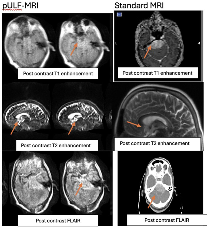
Brain tumors represent a significant burden, particularly in low- and middle-income countries (LMICs) where access to neuroimaging techniques is often limited. Conventional MRI machines are expensive and bulky, posing a significant challenge in the diagnosis and treatment of brain tumors in LMICs. However, an emerging technology, ultra-low field magnetic resonance imaging (pULF-MRI), has the potential to address this limitation. This study aimed to evaluate the feasibility and effectiveness of post-contrast enhancement in a pULF-MRI scanner for brain tumor imaging in LMICs. A single case study was conducted, and post-contrast enhancement was successfully achieved, revealing the presence of a tumor which was subsequently confirmed on biopsy. To our knowledge, this is the first study to demonstrate the feasibility of post-contrast enhancement in a pULF-MRI scanner for brain tumor imaging. This technology has the potential to significantly improve access to neuroimaging in LMICs, leading to earlier diagnosis and more effective treatment of brain tumors. These promising results suggest that further studies are warranted to explore the potential of pULF-MRI for large-scale screening and diagnosis of brain tumors in LMICs. This can provide a future roadmap for neuroimaging in LMICs, providing a cost-effective and accessible way to diagnose and treat brain tumors, leading to improved healthcare outcomes with a further prospective clinical trial.

 求助内容:
求助内容: 应助结果提醒方式:


