Hinnerk Schulz-Hildebrandt, Michael Wang-Evers, Naja Meyer-Schell, Daniel Karasik, Malte J Casper, Tim Eixmann, Felix Hilge, Reginald Birngruber, Dieter Manstein, Gereon Hüttmann
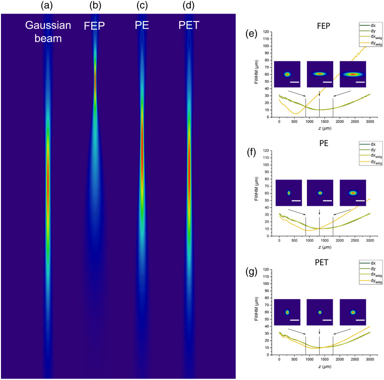
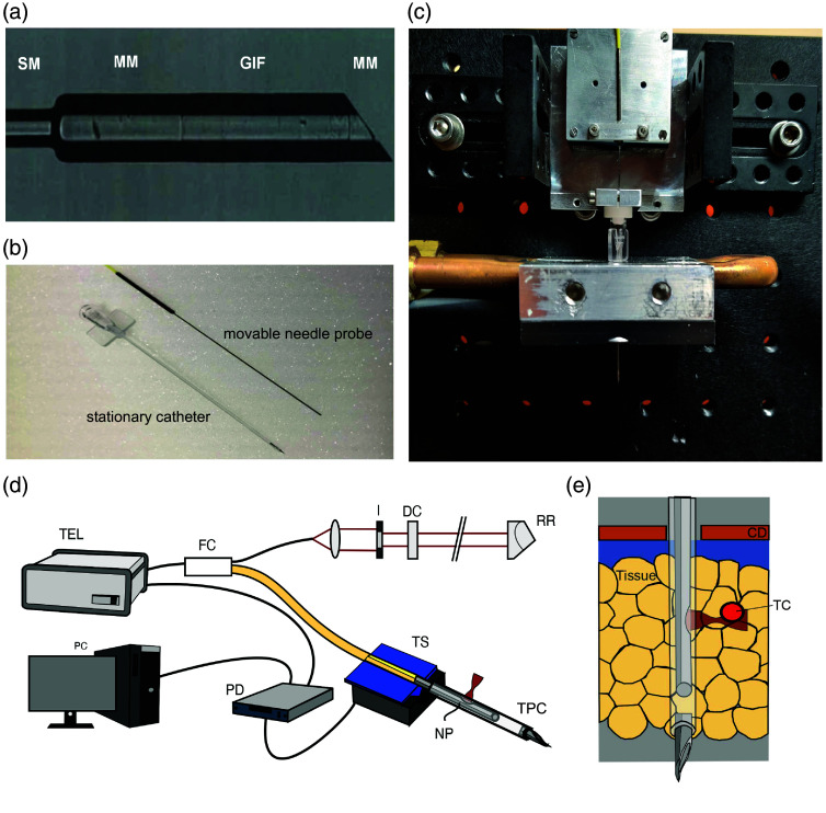
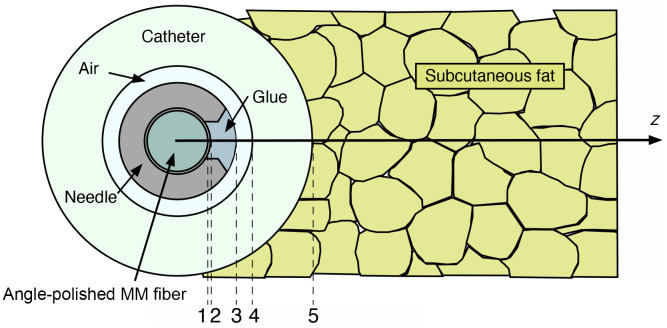
{"title":"Optical coherence tomography needle probe for real-time visualization of temperature-induced phase changes within subcutaneous fatty tissue.","authors":"Hinnerk Schulz-Hildebrandt, Michael Wang-Evers, Naja Meyer-Schell, Daniel Karasik, Malte J Casper, Tim Eixmann, Felix Hilge, Reginald Birngruber, Dieter Manstein, Gereon Hüttmann","doi":"10.1117/1.JBO.30.3.035002","DOIUrl":null,"url":null,"abstract":"<p><p><b>Significance</b>: Selective cryolipolysis is a widely used aesthetic procedure that cools subcutaneous adipose tissue to temperatures as low as <math><mrow><mo>-</mo> <mn>11</mn> <mo>°</mo> <mi>C</mi></mrow> </math> to induce fat cell destruction. However, real-time monitoring techniques are lacking, limiting the ability to optimize safety and efficacy. Traditional imaging methods either fail to provide adequate penetration depth or lack the resolution necessary for visualizing subcutaneous fatty tissue dynamics. <b>Aim</b>: This paper aims to demonstrate that an optical coherence tomography (OCT) needle probe can be used for real-time observation of temperature-induced changes in subcutaneous fatty tissue, potentially enhancing the assessment and optimization of cryolipolysis procedures. <b>Approach</b>: We developed a side-viewing OCT-based needle probe designed for subcutaneous imaging. The probe consists of a fiber-optic system encased in a transparent, biocompatible polymer catheter with an outer diameter of <math><mrow><mn>900</mn> <mtext> </mtext> <mi>μ</mi> <mi>m</mi></mrow> </math> . A 49-degree angled fiber enables imaging, while a piezoelectric scanning system moves the fiber transversely within the catheter. The probe achieves a lateral resolution of <math><mrow><mo><</mo> <mn>15</mn> <mtext> </mtext> <mi>μ</mi> <mi>m</mi></mrow> </math> , a working distance of <math><mrow><mn>600</mn> <mtext> </mtext> <mi>μ</mi> <mi>m</mi></mrow> </math> , and a lateral field of view dictated by the scanning system length. OCT imaging was performed on porcine skin with a subcutaneous fat layer >3 cm thick during controlled heating and cooling. <b>Results</b>: OCT imaging revealed increased optical scattering in subcutaneous fatty tissue during cooling, corresponding to the phase transition from liquid to solid. This effect was reversible upon warming, indicating that OCT can dynamically monitor adipocyte crystallization in real time. The observed transition temperatures varied, likely due to differences in lipid composition. <b>Conclusions</b>: OCT-based needle imaging enables direct, high-resolution visualization of adipocyte crystallization, offering a potential tool for optimizing selective cryolipolysis treatments. This technology could improve safety and efficacy by providing real-time feedback on tissue response, facilitating a better understanding of the cooling-induced fat reduction process.</p>","PeriodicalId":15264,"journal":{"name":"Journal of Biomedical Optics","volume":"30 3","pages":"035002"},"PeriodicalIF":2.9000,"publicationDate":"2025-03-01","publicationTypes":"Journal Article","fieldsOfStudy":null,"isOpenAccess":false,"openAccessPdf":"https://www.ncbi.nlm.nih.gov/pmc/articles/PMC11895999/pdf/","citationCount":"0","resultStr":null,"platform":"Semanticscholar","paperid":null,"PeriodicalName":"Journal of Biomedical Optics","FirstCategoryId":"3","ListUrlMain":"https://doi.org/10.1117/1.JBO.30.3.035002","RegionNum":3,"RegionCategory":"医学","ArticlePicture":[],"TitleCN":null,"AbstractTextCN":null,"PMCID":null,"EPubDate":"2025/3/11 0:00:00","PubModel":"Epub","JCR":"Q2","JCRName":"BIOCHEMICAL RESEARCH METHODS","Score":null,"Total":0}
引用次数: 0
Abstract
Significance: Selective cryolipolysis is a widely used aesthetic procedure that cools subcutaneous adipose tissue to temperatures as low as to induce fat cell destruction. However, real-time monitoring techniques are lacking, limiting the ability to optimize safety and efficacy. Traditional imaging methods either fail to provide adequate penetration depth or lack the resolution necessary for visualizing subcutaneous fatty tissue dynamics. Aim: This paper aims to demonstrate that an optical coherence tomography (OCT) needle probe can be used for real-time observation of temperature-induced changes in subcutaneous fatty tissue, potentially enhancing the assessment and optimization of cryolipolysis procedures. Approach: We developed a side-viewing OCT-based needle probe designed for subcutaneous imaging. The probe consists of a fiber-optic system encased in a transparent, biocompatible polymer catheter with an outer diameter of . A 49-degree angled fiber enables imaging, while a piezoelectric scanning system moves the fiber transversely within the catheter. The probe achieves a lateral resolution of , a working distance of , and a lateral field of view dictated by the scanning system length. OCT imaging was performed on porcine skin with a subcutaneous fat layer >3 cm thick during controlled heating and cooling. Results: OCT imaging revealed increased optical scattering in subcutaneous fatty tissue during cooling, corresponding to the phase transition from liquid to solid. This effect was reversible upon warming, indicating that OCT can dynamically monitor adipocyte crystallization in real time. The observed transition temperatures varied, likely due to differences in lipid composition. Conclusions: OCT-based needle imaging enables direct, high-resolution visualization of adipocyte crystallization, offering a potential tool for optimizing selective cryolipolysis treatments. This technology could improve safety and efficacy by providing real-time feedback on tissue response, facilitating a better understanding of the cooling-induced fat reduction process.
期刊介绍:
The Journal of Biomedical Optics publishes peer-reviewed papers on the use of modern optical technology for improved health care and biomedical research.

 求助内容:
求助内容: 应助结果提醒方式:


