{"title":"Quantitative Detection of Orthotopic Liver Cancer in Mice Using Indocyanine Green and Dynamic Diffuse Fluorescence Tomography Imaging","authors":"Zhuanxia He, Limin Zhang, Zishuo Li, Xiujun Gao, Yanqi Zhang, Feng Gao","doi":"10.1002/jbio.70003","DOIUrl":null,"url":null,"abstract":"<div>\n \n <p>Orthotopic tumor model has become an essential tool for studying drug biodistribution and tumor progression over time owing to the rapid development of in vivo imaging and immunological science. Dynamic diffuse fluorescence tomography (DFT) is a promising imaging modality that can map the three-dimensional distribution of a fluorophore within the object and capture the metabolic parameters of fluorophores in vivo. It has been widely applied in tumor detection, drug development, and efficacy evaluation. To detect orthotopic liver tumors, we combined indocyanine green (ICG) and a DFT system to perform fluorescence imaging and quantitative analysis for orthotopic liver tumors in mice. The orthotopic liver models were first established, and the liver fluorescence yields were detected in pre- and post-cancerous liver using the DFT system. The results showed that there was higher liver uptake and prolonged retention in orthotopic tumor liver compared to normal liver. Furthermore, the pharmacokinetic parameters suggested the uptake coefficient of tumor liver was above twice that of normal liver, while the excretion rates were similar. Additionally, to intuitively assess tumor occurrence, we propose using normalized fluorescence ratios of 5 and maximum fluorescence values of 0.01 mm<sup>−1</sup> as evaluation criteria. The study demonstrates DFT imaging is a promising tool in orthotopic tumor model detection and drug or agent metabolic evaluation.</p>\n </div>","PeriodicalId":184,"journal":{"name":"Journal of Biophotonics","volume":"18 6","pages":""},"PeriodicalIF":2.0000,"publicationDate":"2025-03-11","publicationTypes":"Journal Article","fieldsOfStudy":null,"isOpenAccess":false,"openAccessPdf":"","citationCount":"0","resultStr":null,"platform":"Semanticscholar","paperid":null,"PeriodicalName":"Journal of Biophotonics","FirstCategoryId":"101","ListUrlMain":"https://onlinelibrary.wiley.com/doi/10.1002/jbio.70003","RegionNum":3,"RegionCategory":"物理与天体物理","ArticlePicture":[],"TitleCN":null,"AbstractTextCN":null,"PMCID":null,"EPubDate":"","PubModel":"","JCR":"Q3","JCRName":"BIOCHEMICAL RESEARCH METHODS","Score":null,"Total":0}
引用次数: 0
Abstract
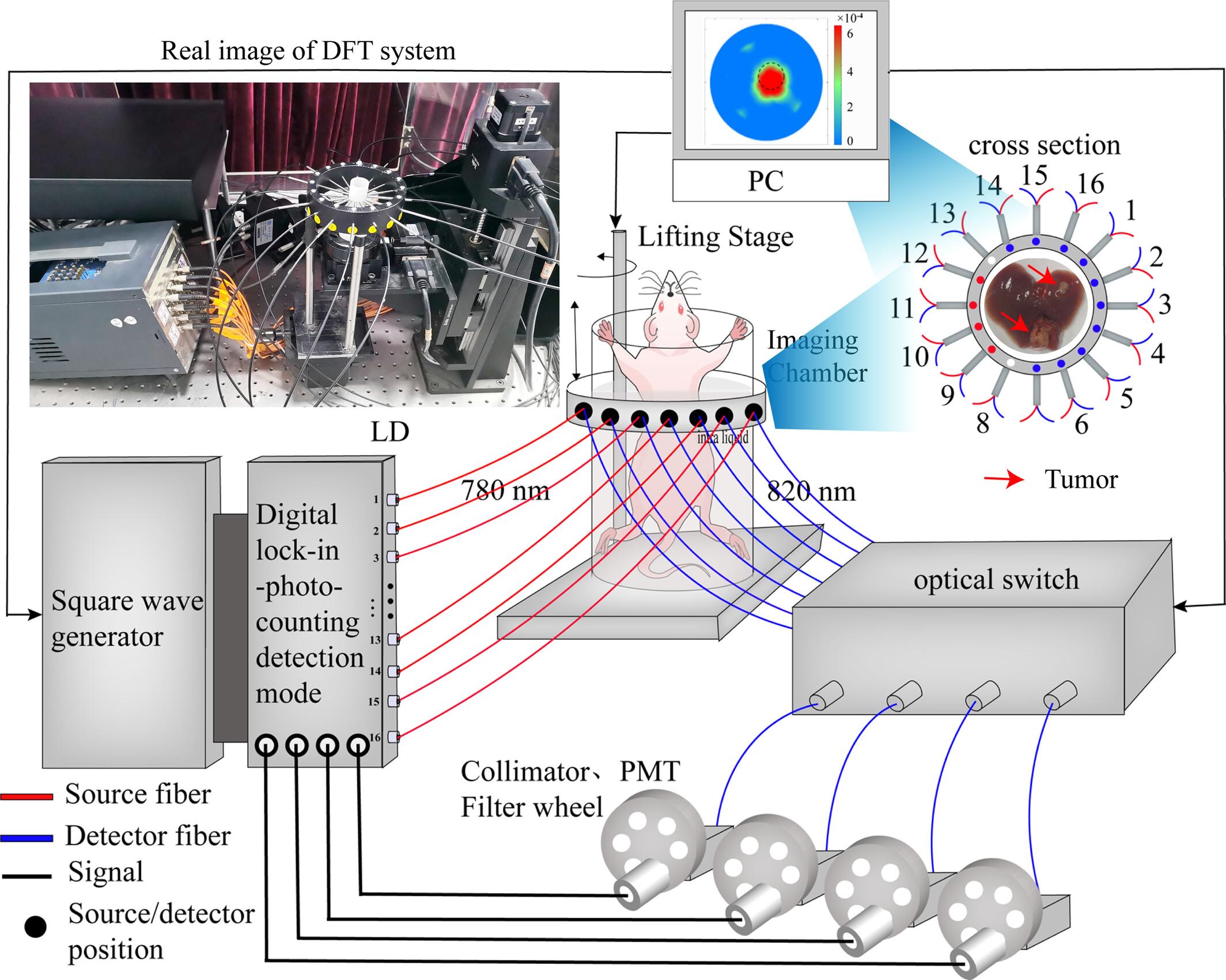
Orthotopic tumor model has become an essential tool for studying drug biodistribution and tumor progression over time owing to the rapid development of in vivo imaging and immunological science. Dynamic diffuse fluorescence tomography (DFT) is a promising imaging modality that can map the three-dimensional distribution of a fluorophore within the object and capture the metabolic parameters of fluorophores in vivo. It has been widely applied in tumor detection, drug development, and efficacy evaluation. To detect orthotopic liver tumors, we combined indocyanine green (ICG) and a DFT system to perform fluorescence imaging and quantitative analysis for orthotopic liver tumors in mice. The orthotopic liver models were first established, and the liver fluorescence yields were detected in pre- and post-cancerous liver using the DFT system. The results showed that there was higher liver uptake and prolonged retention in orthotopic tumor liver compared to normal liver. Furthermore, the pharmacokinetic parameters suggested the uptake coefficient of tumor liver was above twice that of normal liver, while the excretion rates were similar. Additionally, to intuitively assess tumor occurrence, we propose using normalized fluorescence ratios of 5 and maximum fluorescence values of 0.01 mm−1 as evaluation criteria. The study demonstrates DFT imaging is a promising tool in orthotopic tumor model detection and drug or agent metabolic evaluation.
期刊介绍:
The first international journal dedicated to publishing reviews and original articles from this exciting field, the Journal of Biophotonics covers the broad range of research on interactions between light and biological material. The journal offers a platform where the physicist communicates with the biologist and where the clinical practitioner learns about the latest tools for the diagnosis of diseases. As such, the journal is highly interdisciplinary, publishing cutting edge research in the fields of life sciences, medicine, physics, chemistry, and engineering. The coverage extends from fundamental research to specific developments, while also including the latest applications.
 求助内容:
求助内容: 应助结果提醒方式:


