Trabecular-bone mimicking osteoconductive collagen scaffolds: an optimized 3D printing approach using freeform reversible embedding of suspended hydrogels.
IF 3.1 Q1 RADIOLOGY, NUCLEAR MEDICINE & MEDICAL IMAGING
Michael G Kontakis, Marie Moulin, Brittmarie Andersson, Norein Norein, Ayan Samanta, Christina Stelzl, Adam Engberg, Anna Diez-Escudero, Johan Kreuger, Nils P Hailer
{"title":"Trabecular-bone mimicking osteoconductive collagen scaffolds: an optimized 3D printing approach using freeform reversible embedding of suspended hydrogels.","authors":"Michael G Kontakis, Marie Moulin, Brittmarie Andersson, Norein Norein, Ayan Samanta, Christina Stelzl, Adam Engberg, Anna Diez-Escudero, Johan Kreuger, Nils P Hailer","doi":"10.1186/s41205-025-00255-0","DOIUrl":null,"url":null,"abstract":"<p><strong>Background: </strong>Technological constraints limit 3D printing of collagen structures with complex trabecular shapes. However, the Freeform Reversible Embedding of Suspended Hydrogels (FRESH) method may allow for precise 3D printing of porous collagen scaffolds that carry the potential for repairing critical size bone defects.</p><p><strong>Methods: </strong>Collagen type I scaffolds mimicking trabecular bone were fabricated through FRESH 3D printing and compared either with 2D collagen coatings or with 3D-printed polyethylene glycol diacrylate (PEGDA) scaffolds. The porosity of the printed scaffolds was visualized by confocal microscopy, the surface geometry of the scaffolds was investigated by scanning electron microscopy (SEM), and their mechanical properties were assessed with a rheometer. The osteoconductive properties of the different scaffolds were evaluated for up to four weeks by seeding and propagation of primary human osteoblasts (hOBs) or SaOS-2 cells. Intracellular alkaline phosphatase (ALP) and lactate dehydrogenase (LDH) activities were measured, and cells colonizing scaffolds were stained for osteocalcin (OCN).</p><p><strong>Results: </strong>The FRESH technique enables printing of constructs at the millimetre scale using highly concentrated collagen, and the creation of stable trabecular structures that can support the growth osteogenic cells. FRESH-printed collagen scaffolds displayed an intricate and fibrous 3D network, as visualized by SEM, whereas the PEGDA scaffolds had a smooth surface. Amplitude sweep analyses revealed that the collagen scaffolds exhibited predominantly elastic behaviour, as indicated by higher storage modulus values relative to loss modulus values, while the degradation rate of collagen scaffolds was greater than PEGDA. The osteoconductive properties of collagen scaffolds were similar to those of PEGDA scaffolds but superior to 2D collagen, as verified by cell culture followed by analysis of ALP/LDH activity and OCN immunostaining.</p><p><strong>Conclusions: </strong>Our findings suggest that FRESH-printed collagen scaffolds exhibit favourable mechanical, degradation and osteoconductive properties, potentially outperforming synthetic polymers such as PEGDA in bone tissue engineering applications.</p>","PeriodicalId":72036,"journal":{"name":"3D printing in medicine","volume":"11 1","pages":"11"},"PeriodicalIF":3.1000,"publicationDate":"2025-03-11","publicationTypes":"Journal Article","fieldsOfStudy":null,"isOpenAccess":false,"openAccessPdf":"https://www.ncbi.nlm.nih.gov/pmc/articles/PMC11895158/pdf/","citationCount":"0","resultStr":null,"platform":"Semanticscholar","paperid":null,"PeriodicalName":"3D printing in medicine","FirstCategoryId":"1085","ListUrlMain":"https://doi.org/10.1186/s41205-025-00255-0","RegionNum":0,"RegionCategory":null,"ArticlePicture":[],"TitleCN":null,"AbstractTextCN":null,"PMCID":null,"EPubDate":"","PubModel":"","JCR":"Q1","JCRName":"RADIOLOGY, NUCLEAR MEDICINE & MEDICAL IMAGING","Score":null,"Total":0}
引用次数: 0
Abstract
Background: Technological constraints limit 3D printing of collagen structures with complex trabecular shapes. However, the Freeform Reversible Embedding of Suspended Hydrogels (FRESH) method may allow for precise 3D printing of porous collagen scaffolds that carry the potential for repairing critical size bone defects.
Methods: Collagen type I scaffolds mimicking trabecular bone were fabricated through FRESH 3D printing and compared either with 2D collagen coatings or with 3D-printed polyethylene glycol diacrylate (PEGDA) scaffolds. The porosity of the printed scaffolds was visualized by confocal microscopy, the surface geometry of the scaffolds was investigated by scanning electron microscopy (SEM), and their mechanical properties were assessed with a rheometer. The osteoconductive properties of the different scaffolds were evaluated for up to four weeks by seeding and propagation of primary human osteoblasts (hOBs) or SaOS-2 cells. Intracellular alkaline phosphatase (ALP) and lactate dehydrogenase (LDH) activities were measured, and cells colonizing scaffolds were stained for osteocalcin (OCN).
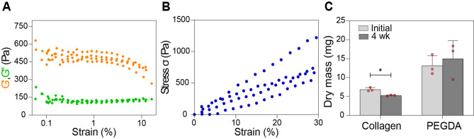
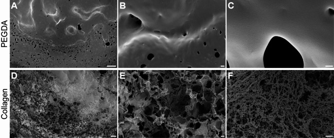
Results: The FRESH technique enables printing of constructs at the millimetre scale using highly concentrated collagen, and the creation of stable trabecular structures that can support the growth osteogenic cells. FRESH-printed collagen scaffolds displayed an intricate and fibrous 3D network, as visualized by SEM, whereas the PEGDA scaffolds had a smooth surface. Amplitude sweep analyses revealed that the collagen scaffolds exhibited predominantly elastic behaviour, as indicated by higher storage modulus values relative to loss modulus values, while the degradation rate of collagen scaffolds was greater than PEGDA. The osteoconductive properties of collagen scaffolds were similar to those of PEGDA scaffolds but superior to 2D collagen, as verified by cell culture followed by analysis of ALP/LDH activity and OCN immunostaining.
Conclusions: Our findings suggest that FRESH-printed collagen scaffolds exhibit favourable mechanical, degradation and osteoconductive properties, potentially outperforming synthetic polymers such as PEGDA in bone tissue engineering applications.

 求助内容:
求助内容: 应助结果提醒方式:


