Seon Hui Kim, Hye Yoon Chung, MinGi Kim, Seung Woo Kim, Ha Young Shin
{"title":"Development and Application of a Cell-Based Assay for Detecting Anti-Agrin Antibodies Associated With Myasthenia Gravis.","authors":"Seon Hui Kim, Hye Yoon Chung, MinGi Kim, Seung Woo Kim, Ha Young Shin","doi":"10.3988/jcn.2024.0413","DOIUrl":null,"url":null,"abstract":"<p><strong>Background and purpose: </strong>Anti-agrin antibodies (agrin Abs) have recently been identified in patients with myasthenia gravis (MG), sometimes in conjunction with antibodies (Abs) to the acetylcholine receptor (AChR), muscle-specific tyrosine kinase (MuSK), or low-density lipoprotein receptor-related protein 4. This study aimed to develop an in-house cell-based assay (CBA) for detecting agrin Abs, and to test its application to serum samples collected from individuals diagnosed with MG.</p><p><strong>Methods: </strong>Agrin complementary DNA as cloned into a pCMV6-AC-GFP vector, which was subsequently transfected into human embryonic kidney 293T (HEK293T) cells. Transfected HEK293T cells were incubated with patient serum and antihuman immunoglobulin G Ab conjugated with a red fluorescent dye. Agrin Ab levels were measured using the CBA in 389 serum samples: 340 from patients with MG, 36 from patients with other neuromuscular diseases, and 13 from healthy controls. The presence of agrin Ab was determined based on the fluorescence intensity and colocalization using fluorescence microscopy.</p><p><strong>Results: </strong>The expression levels of agrin mRNA and protein in transfected HEK293T cells were confirmed using the reverse-transcription polymerase chain reaction and Western blotting, respectively. Agrin expression in cells was further confirmed by immunocytochemistry. Two (0.6%) of the 340 patients with MG tested positive for agrin Ab: 1 of 191 AChR-positive patients and 1 of 54 MuSK-positive patients.</p><p><strong>Conclusions: </strong>We have developed and validated a novel CBA for detecting agrin Abs. This CBA was successfully applied to detect agrin Abs in serum samples obtained from individuals with MG.</p>","PeriodicalId":15432,"journal":{"name":"Journal of Clinical Neurology","volume":"21 2","pages":"105-112"},"PeriodicalIF":3.1000,"publicationDate":"2025-03-01","publicationTypes":"Journal Article","fieldsOfStudy":null,"isOpenAccess":false,"openAccessPdf":"https://www.ncbi.nlm.nih.gov/pmc/articles/PMC11896744/pdf/","citationCount":"0","resultStr":null,"platform":"Semanticscholar","paperid":null,"PeriodicalName":"Journal of Clinical Neurology","FirstCategoryId":"3","ListUrlMain":"https://doi.org/10.3988/jcn.2024.0413","RegionNum":3,"RegionCategory":"医学","ArticlePicture":[],"TitleCN":null,"AbstractTextCN":null,"PMCID":null,"EPubDate":"","PubModel":"","JCR":"Q2","JCRName":"CLINICAL NEUROLOGY","Score":null,"Total":0}
引用次数: 0
Abstract
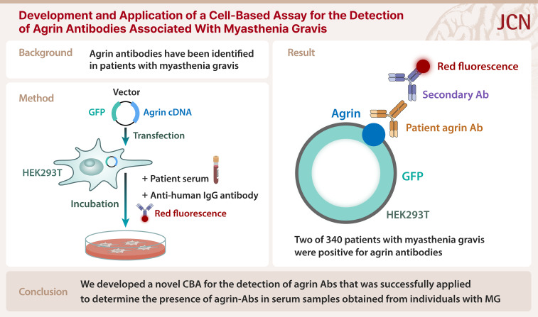
Background and purpose: Anti-agrin antibodies (agrin Abs) have recently been identified in patients with myasthenia gravis (MG), sometimes in conjunction with antibodies (Abs) to the acetylcholine receptor (AChR), muscle-specific tyrosine kinase (MuSK), or low-density lipoprotein receptor-related protein 4. This study aimed to develop an in-house cell-based assay (CBA) for detecting agrin Abs, and to test its application to serum samples collected from individuals diagnosed with MG.
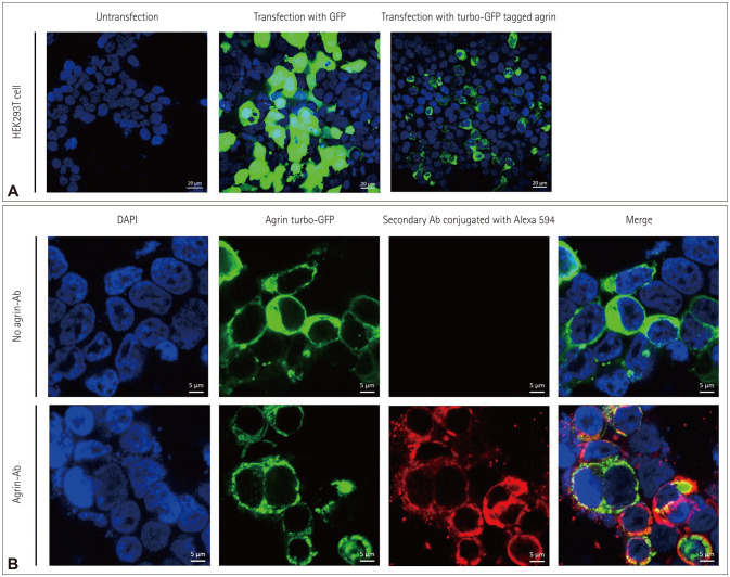
Methods: Agrin complementary DNA as cloned into a pCMV6-AC-GFP vector, which was subsequently transfected into human embryonic kidney 293T (HEK293T) cells. Transfected HEK293T cells were incubated with patient serum and antihuman immunoglobulin G Ab conjugated with a red fluorescent dye. Agrin Ab levels were measured using the CBA in 389 serum samples: 340 from patients with MG, 36 from patients with other neuromuscular diseases, and 13 from healthy controls. The presence of agrin Ab was determined based on the fluorescence intensity and colocalization using fluorescence microscopy.
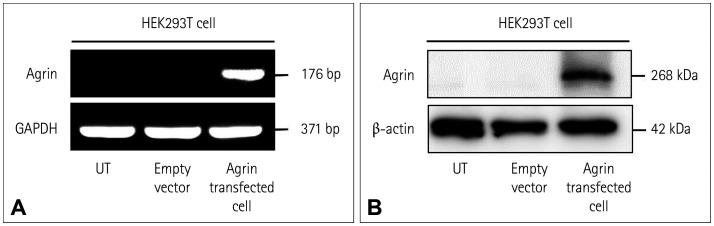
Results: The expression levels of agrin mRNA and protein in transfected HEK293T cells were confirmed using the reverse-transcription polymerase chain reaction and Western blotting, respectively. Agrin expression in cells was further confirmed by immunocytochemistry. Two (0.6%) of the 340 patients with MG tested positive for agrin Ab: 1 of 191 AChR-positive patients and 1 of 54 MuSK-positive patients.
Conclusions: We have developed and validated a novel CBA for detecting agrin Abs. This CBA was successfully applied to detect agrin Abs in serum samples obtained from individuals with MG.
期刊介绍:
The JCN aims to publish the cutting-edge research from around the world. The JCN covers clinical and translational research for physicians and researchers in the field of neurology. Encompassing the entire neurological diseases, our main focus is on the common disorders including stroke, epilepsy, Parkinson''s disease, dementia, multiple sclerosis, headache, and peripheral neuropathy. Any authors affiliated with an accredited biomedical institution may submit manuscripts of original articles, review articles, and letters to the editor. The JCN will allow clinical neurologists to enrich their knowledge of patient management, education, and clinical or experimental research, and hence their professionalism.
 求助内容:
求助内容: 应助结果提醒方式:


