Fatemeh Eslami, Hamidreza Ghasemibasir, Sara Alipour, Ramin Mansouri
{"title":"Evaluating the Diagnostic Accuracy of Impression Cytology for Conjunctival Lesions: A Comparative Study with Histopathology.","authors":"Fatemeh Eslami, Hamidreza Ghasemibasir, Sara Alipour, Ramin Mansouri","doi":"10.30699/ijp.2024.2025786.3284","DOIUrl":null,"url":null,"abstract":"<p><strong>Background & objective: </strong>Conjunctival lesions have a wide range of histological manifestations that are difficult to distinguish clinically. The gold standard for diagnosis of these lesions is the pathological examination, a costly and invasive procedure that may also adversely affect patients. Therefore, clinical researchers seek less invasive, inexpensive, and easier methods to detect conjunctival lesions. This study aims to compare the accuracy of impression cytology with pathology in patients referred to Farshchian Hospital in Iran.</p><p><strong>Methods: </strong>In this descriptive/cross-sectional study, 64 patients with conjunctival lesions were selected from patients referred to Farshchian Hospital in Hamedan in 2021. A cytology specimen was obtained from the patients and sent to the laboratory. The diagnostic accuracy of this method was compared with pathological results (gold standard). Data were analyzed by SPSS 16 software.</p><p><strong>Results: </strong>The mean age of patients was 54.47 ±16.94 years; 70.3% were male, and 29.7% were female. In the cytologic and pathologic examination, 28.1% of the specimens showed dysplasia, and 71.9% were non-dysplastic. The sensitivity, specificity, positive predictive value, negative predictive value, and accuracy of the cytologic impression compared to the pathologic methods were 91.30%, 77.78%, 91.30%, 77.78%, and 87.5%, respectively. A positive and significant correlation was observed between pathological and cytological diagnosis scores (r=0.825, P-value<0.001).</p><p><strong>Conclusion: </strong>In conjunctival lesions, impression cytology may be a relatively accurate and sensitive procedure that can distinguish dysplastic from non-dysplastic conjunctival lesions.</p>","PeriodicalId":38900,"journal":{"name":"Iranian Journal of Pathology","volume":"20 1","pages":"49-57"},"PeriodicalIF":0.0000,"publicationDate":"2025-01-01","publicationTypes":"Journal Article","fieldsOfStudy":null,"isOpenAccess":false,"openAccessPdf":"https://www.ncbi.nlm.nih.gov/pmc/articles/PMC11887644/pdf/","citationCount":"0","resultStr":null,"platform":"Semanticscholar","paperid":null,"PeriodicalName":"Iranian Journal of Pathology","FirstCategoryId":"1085","ListUrlMain":"https://doi.org/10.30699/ijp.2024.2025786.3284","RegionNum":0,"RegionCategory":null,"ArticlePicture":[],"TitleCN":null,"AbstractTextCN":null,"PMCID":null,"EPubDate":"2025/1/10 0:00:00","PubModel":"Epub","JCR":"Q3","JCRName":"Medicine","Score":null,"Total":0}
引用次数: 0
Abstract
Background & objective: Conjunctival lesions have a wide range of histological manifestations that are difficult to distinguish clinically. The gold standard for diagnosis of these lesions is the pathological examination, a costly and invasive procedure that may also adversely affect patients. Therefore, clinical researchers seek less invasive, inexpensive, and easier methods to detect conjunctival lesions. This study aims to compare the accuracy of impression cytology with pathology in patients referred to Farshchian Hospital in Iran.
Methods: In this descriptive/cross-sectional study, 64 patients with conjunctival lesions were selected from patients referred to Farshchian Hospital in Hamedan in 2021. A cytology specimen was obtained from the patients and sent to the laboratory. The diagnostic accuracy of this method was compared with pathological results (gold standard). Data were analyzed by SPSS 16 software.
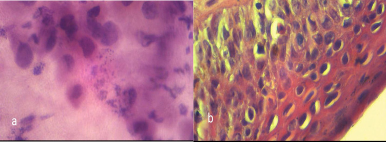
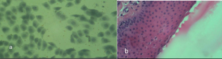
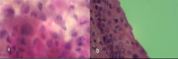
Results: The mean age of patients was 54.47 ±16.94 years; 70.3% were male, and 29.7% were female. In the cytologic and pathologic examination, 28.1% of the specimens showed dysplasia, and 71.9% were non-dysplastic. The sensitivity, specificity, positive predictive value, negative predictive value, and accuracy of the cytologic impression compared to the pathologic methods were 91.30%, 77.78%, 91.30%, 77.78%, and 87.5%, respectively. A positive and significant correlation was observed between pathological and cytological diagnosis scores (r=0.825, P-value<0.001).
Conclusion: In conjunctival lesions, impression cytology may be a relatively accurate and sensitive procedure that can distinguish dysplastic from non-dysplastic conjunctival lesions.

 求助内容:
求助内容: 应助结果提醒方式:


